
System and method for automatic color segmentation and minimum significant response for measurement of fractional localized intensity of cellular compartments

a fractional localized intensity and color segmentation technology, applied in the field of biological material assays, can solve the problems of compromising the efficacy of candidate drugs, unable to meet the requirements of cost-effective and controversial animal testing, and complex real cell responses, etc., to achieve minimum significant response, high throughput screening, and economic advantages

Inactive Publication Date: 2013-01-03
VALA SCI
View PDF1 Cites 4 Cited by
  • Summary
  • Abstract
  • Description
  • Claims
  • Application Information

AI Technical Summary

Benefits of technology

The invention is a system and method that automates the process of measuring the intensity of cellular compartments. It uses advanced techniques to accurately analyze color and minimize errors. This invention has the technical advantage of making the measurement process faster, more efficient, and more reliable.

Problems solved by technology

Multi-parameter cell assays, where a response is measured by multiplexed reporter molecules, as well as morphological criteria, have been limited by the labor-intensive nature of imaging and analyzing subcellular details.
If the signaling pathway used also led to cell death, the efficacy of the candidate drug would be compromised and would fail in costly and controversial animal testing.
The complexity of real cell responses also leads to heterogeneity between cells, even in a cloned cell line, depending on other factors such as cell cycle progression.
If an average response from all the cells in a well is measured, it may fall below the threshold for detection and result in a false negative: an effective drug is overlooked.

Method used


Image

  • System and method for automatic color segmentation and minimum significant response for measurement of fractional localized intensity of cellular compartments
  • System and method for automatic color segmentation and minimum significant response for measurement of fractional localized intensity of cellular compartments
  • System and method for automatic color segmentation and minimum significant response for measurement of fractional localized intensity of cellular compartments

Examples


Embodiment Construction

Fractional Localized Intensity of Cellular Compartments (FLIC)

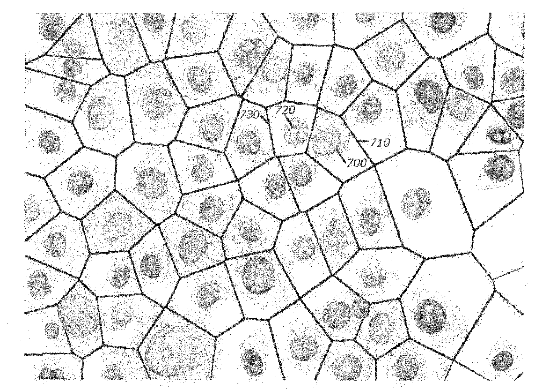
[0044]Development of multicompartment models for cellular translocation events: Many potentially important molecular targets are regulated not only in their expression levels, but also by their subcellular or spatial localization. In the post-genomics era, illuminating the function of genes is rapidly generating new data and overthrowing old dogma. The prevailing picture of the cell is no longer a suspension of proteins, lipids and ions floating around inside a membrane bag, but involves protein complexes attached to architectural scaffolds or chromatin provided by the cytoskeleton, endoplasmic reticulum, Golgi apparatus, ion channels and membrane pores. Cell surface receptors are oriented within the plasma membrane such that they can bind an extracellular molecule and initiate a response in the cytoplasm. Protein complexes in the cytoplasm can be dissociated following regulated proteolysis to release specific DNA binding...


PUM


Abstract

A system, a method, and a programmed device for measurement of translocational activity among cellular compartments process magnified images of cellular material exposed to an agent by segmenting and compartmentalizing the images and then measuring fractional localized intensity of two or more compartments in the segmented, compartmentalized image. The measured fractional localized intensities are compared to determine translocation of cellular material among the measured components caused by the agent.

Description

RELATED APPLICATIONS[0001]This application is a continuation of U.S. patent application Ser. No. 10 / 507,442, §371(c)(1),(2),(4) date Mar. 15, 2006, which is a U.S. national phase under §371 of PCT Application PCT / US03 / 07968, which claims priority from U.S. Provisional Patent Application 60 / 363,889, filed Mar. 13, 2002.TECHNICAL FIELD[0002]This application concerns assay of biological material by means of high speed, high throughput cytometry using fractionalized localized intensities of subcellular components in magnified images.BACKGROUND ART[0003]Drug discovery screening has historically used simple well-based read-outs in order to handle a high throughput of lead compounds. However, a given assay currently provides only the information that a drug affects some of the cellular processes that result in the response measured; the exact nature of the target for the drug is not indicated. A cell-based assay is a model system designed to identify compounds that interact with a selected...

Claims


Application Information

Patent Timeline
Patent Type & Authority Applications(United States)
IPC IPC(8): G06K9/00C12M1/34C12M1/00C12Q1/02G01N15/14G06T5/00G06T7/00
CPCG01N15/1475G06K9/0014G06T7/0012G06T2207/30024G06T2207/10056G06T2207/10064G06T2207/20148G06T7/0081G06T7/11G06T7/136G06V20/695G01N15/1433
Inventor HUNTER, EDWARD A.INGERMANSON, RANDALL SCOTTCALLAWAY, WILLIAM SCOTT
Owner VALA SCI
Learn More