Eureka AIR delivers breakthrough ideas for toughest innovation challenges, trusted by R&D personnel around the world.

Atrial pressure regulation with control, sensing, monitoring and therapy delivery

a technology of atrial pressure and control, applied in the field of atrial pressure regulation with control, sensing, monitoring and therapy delivery, can solve the problems of increased end-diastolic pressure, few, if any, proven effective treatments, and symptomatic, and achieve the effect of reducing damage to blood components

Active Publication Date: 2012-10-18
CORVIA MEDICAL
View PDF5 Cites 160 Cited by
  • Summary
  • Abstract
  • Description
  • Claims
  • Application Information

AI Technical Summary

Benefits of technology

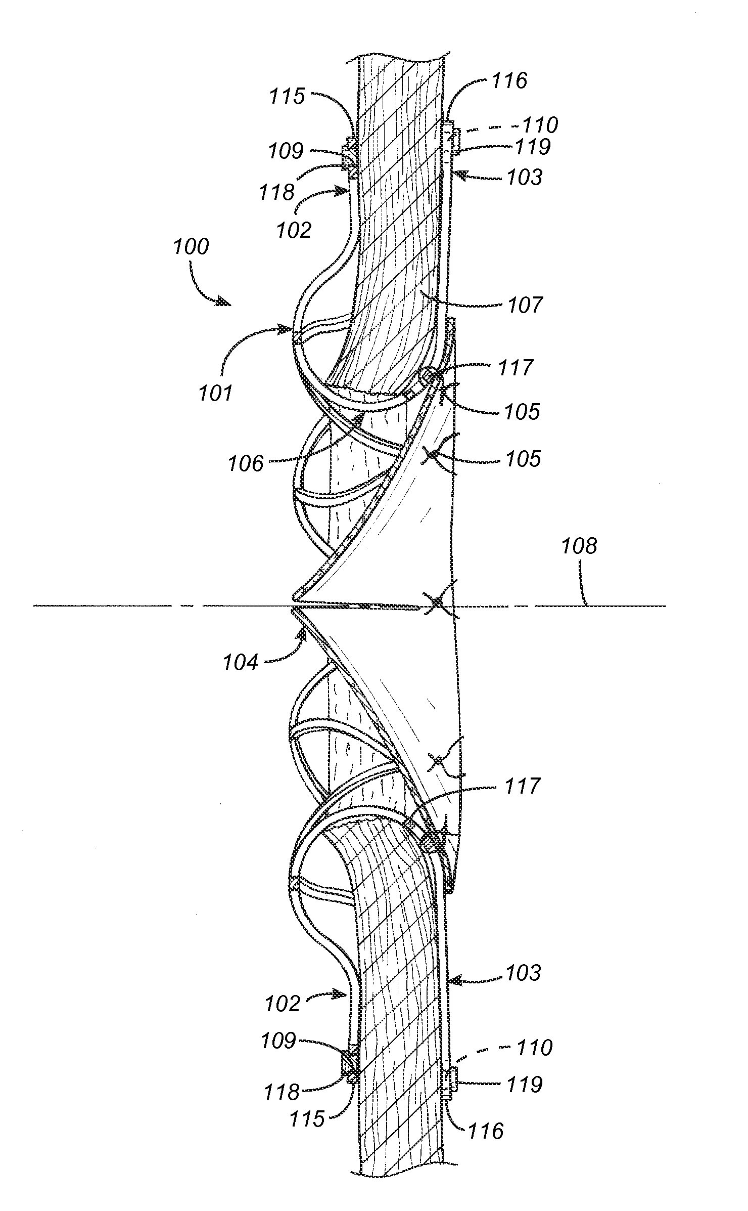
[0022]In embodiments, the body assembly may be constructed from preformed wire braid. The wire braid may be formed from nitinol with a martensite / austenite transition temperature is below 37° C. so it remains in its superelastic, austenitic phase during use. The transition temperature is below about 25+ / −5° C. The wire should have a diameter of at least about 0.0035 in. (about 0.09 mm) with about 2 lbs. of breaking strength at 200 ksi tensile. The wire should have a very smooth surface to reduce thrombogenicity or irritation response from the tissue. The surface finish may be 63 μin RA or better. This surface may be obtained either by mechanical polishing, by electropolishing or a combination. In embodiments, the surface may be cleaned with detergents, acids and / or solvents to remove residual oils or contamination and then controllably passivated to insure minimal corrosion.
[0026]In embodiments, the internal diameter of the finished interatrial pressure vent is larger than about 5 mm to enable adequate venting of the left atrium and minimize damage to blood components from excessive shear stress, but enabling the interatrial pressure vent to stow in a placement catheter of smaller than about 14 F.

Problems solved by technology

Heart failure is a common and potentially lethal condition affecting humans, with sub-optimal clinical outcomes, often resulting in symptoms, morbidity and / or mortality, despite maximal medical treatment.
This condition is characterized by a stiff left ventricle with decreased compliance and impaired relaxation, which leads to increased end-diastolic pressure.
Approximately one third of patients with heart failure have diastolic heart failure and there are very few, if any, proven effective treatments.
In addition to diastolic heart failure, a number of other medical conditions, including systolic dysfunction of the left ventricle and valve disease, can lead to elevated pressures in the left atrium.
Increased left atrial pressure often causes acute or chronic breathlessness amongst other problems.
In addition, a variety of heart conditions can lead to “right heart failure”, which can result in enlargement of the liver (hepatomegaly), fluid accumulation in the abdomen (ascites) and / or swelling of the lower limbs.
As blood backs up into the pulmonary system, fluid leaks into the lungs and causes pulmonary edema.
Blood volume going to the atrium reduces volume of blood going forward into the aorta causing low cardiac output.
Excess blood in the atrium over-fills the ventricle during each cardiac cycle and causes volume overload in the left ventricle.
Heart failure with such symptoms is a common and potentially lethal condition affecting humans, with sub-optimal clinical outcomes often resulting in symptoms, morbidity and / or mortality, despite maximal medical treatment.
This condition is characterized by a stiff left ventricle with decreased compliance and impaired relaxation, which leads to increased end-diastolic pressure.
Approximately one third of patients with heart failure have diastolic heart failure and there are very few, if any, proven effective treatments.
In addition to diastolic heart failure, a number of other medical conditions, including systolic dysfunction of the left ventricle and valve disease, can lead to elevated pressures in the left atrium.
Increased left atrial pressure often causes acute or chronic breathlessness amongst other problems.
In addition, a variety of heart conditions can lead to “right heart failure”, which can result in enlargement of the liver (hepatomegaly), fluid accumulation in the abdomen (ascites) and / or swelling of the lower limbs.

Method used


Image

  • Atrial pressure regulation with control, sensing, monitoring and therapy delivery
  • Atrial pressure regulation with control, sensing, monitoring and therapy delivery
  • Atrial pressure regulation with control, sensing, monitoring and therapy delivery

Examples


embodiment 200

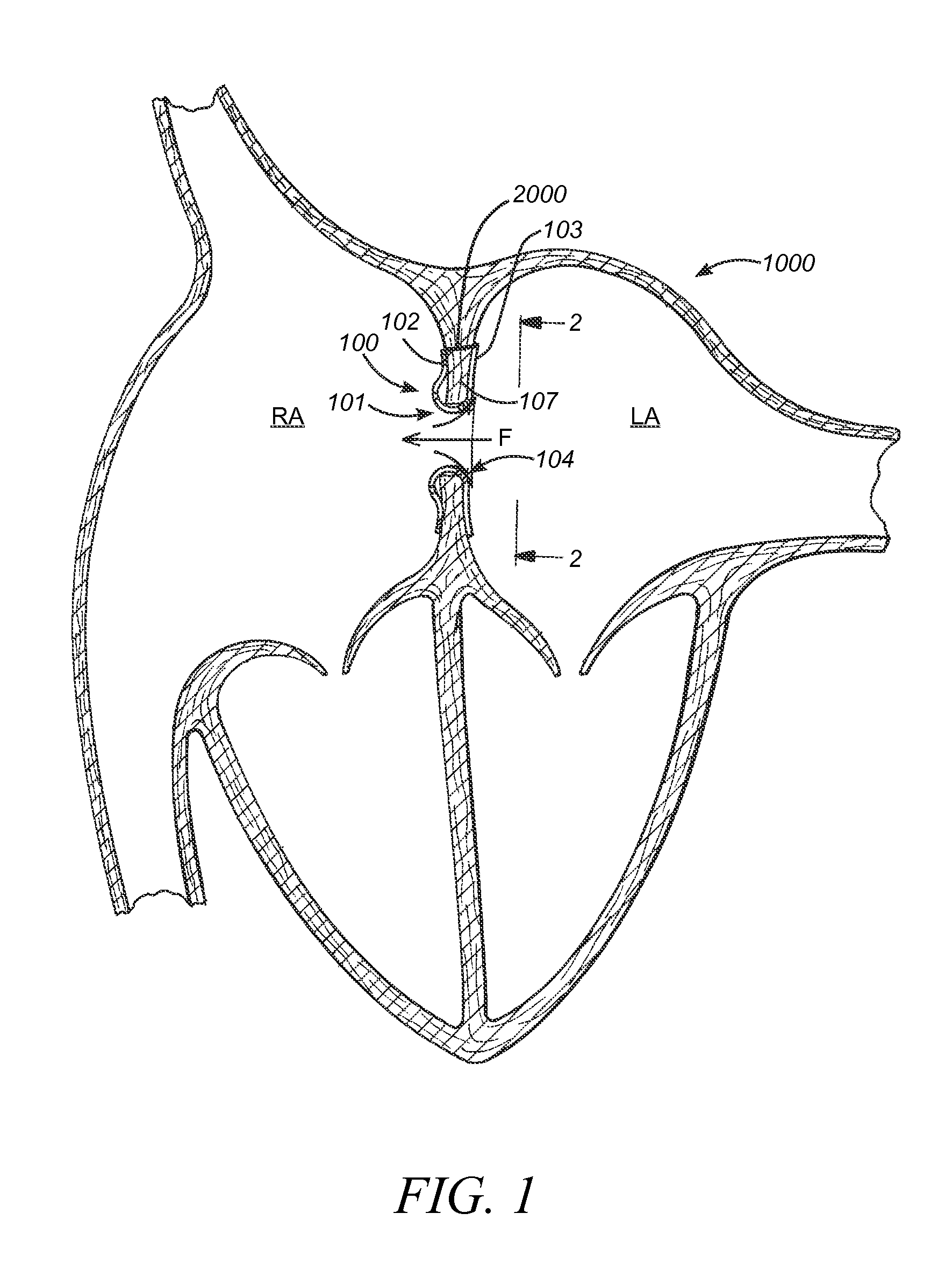
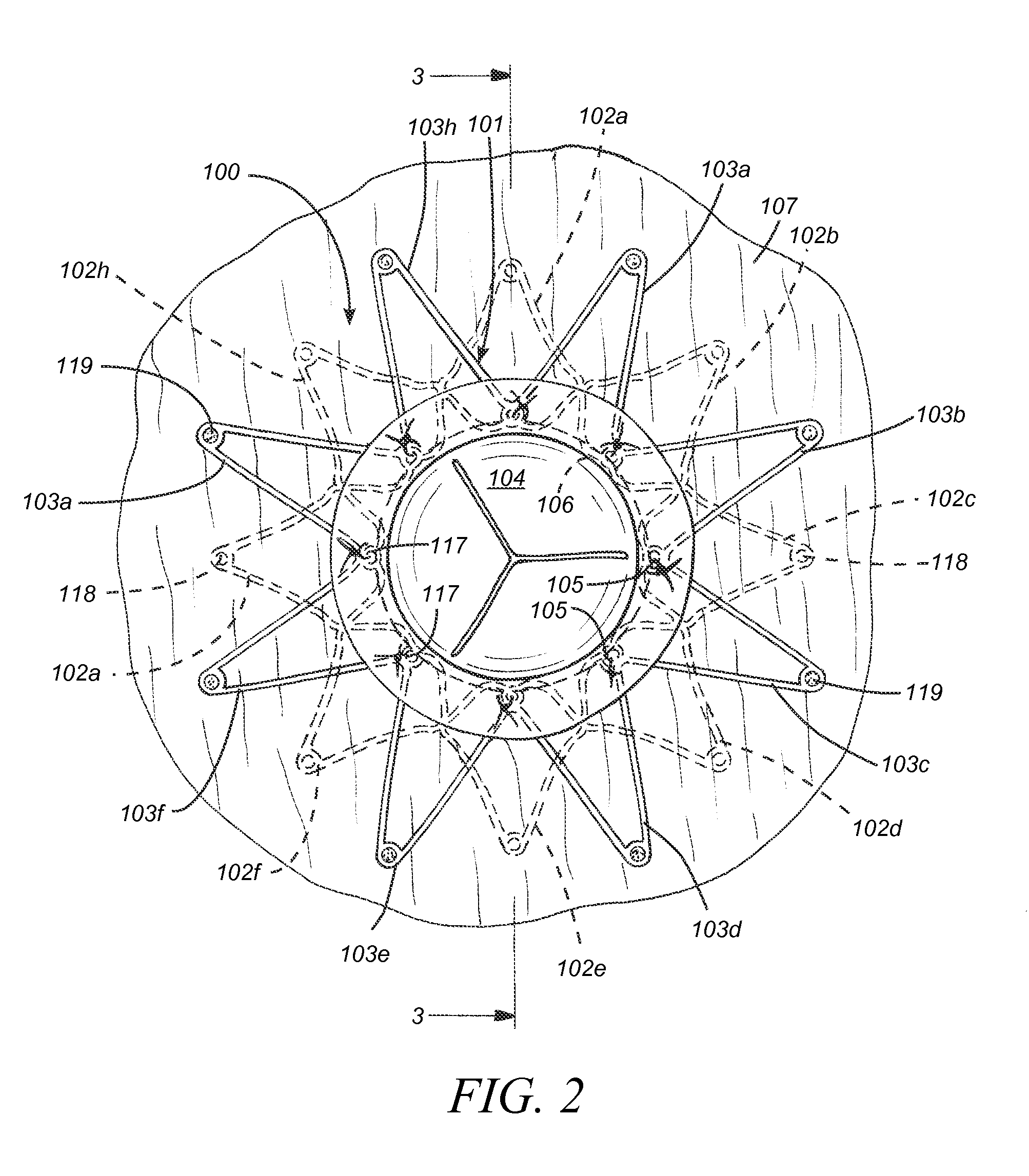
[0142]Referring now to FIG. 8, an interatrial pressure vent 100 is shown in its stowed configuration. In embodiments, the interatrial pressure vent can be collapsed to a substantially cylindrical shape for stowing in a delivery catheter during placement. Flange segments 102a-102h and 103a-103h can be fabricated to be substantially equal in length. The “stowed position” is not meant to apply only to devices having flange segments of equal length but rather to all embodiments of the venting device disclosed herein. Devices having flange segments of varying length and orientation such as those described herein are also designed to stow in substantially the same manner as shown in FIG. 8. In an embodiment 200 seen in FIG. 20, flange segments 202a-202h and 203a-203h are formed on a slanted angle so that, when marker elements are secured to the ends of the flange segments, the flange segments can be stowed into a smaller volume. In embodiments 300 seen in FIG. 21, flange segments 302a-302...

first embodiment

[0180]FIG. 31 depicts a mounting and loading tool useful for placing the prosthesis onto a catheter or other delivery device for delivery in vivo to a patient. In this embodiment, mounting tool 2001 includes a base plate 2002 with orifices 2003 for securing other components as shown with fasteners 2004 and pin 2009. The principal component is a loader body 2014, mounted via the outer two fasteners and orifices as shown. A mounting platform 2023 is mounted in the center of the loader body via the third orifice and pin 2009. Mounting platform 2023 includes a lower orifice 2026 for mounting to the loader body via the middle loader body orifice with pin 2009. Mounting platform 2023 also includes a slotted cam surface 2024. Pivot 2029 mounts to the loader body 2014 via pivot pin 2028 through pivot orifice 2030 and loader body orifice 2016. Pivot 2029 and lever 2031 mount on the left side of loader body 2014 below the side doors 2020, as seen in FIG. 31. Movement of pivot 2029 and lever 2...

third embodiment

[0270]a design for retrieval is depicted in FIGS. 48A and 48B. In this embodiment, prosthesis 151 is very similar to prosthesis 141 above, including retrieval sutures or wires 153 from bores 109 of the right atrium flange apices 102a-h, to a central annular retrieval suture or wire 157. Each retrieval suture or wire 153 is joined to the central retrieval thread 157 at a juncture 155. The junctures may simply be suture tie-offs; alternatively, the junctures could be orifices in central wire 157 for joining retrieval sutures or wires 153. In some embodiments, an additional retrieval suture or wire 147, suitable for non-invasive imaging, may be tied to the central thread at least at one point for grasping by a retrieval device.

[0271]A fourth embodiment of a prosthesis 161 designed for retrieval and redeployment is depicted in FIG. 49. Prosthesis 161 is similar to prosthesis 100a, described above. In the fourth embodiment, there is a retrieval wire or suture 163 secured to each apex 102...


PUM


Abstract

The present disclosure relates to improved capabilities for stabilizing and regulating atrial pressure with a shunt in the atrial septum or a stent in the coronary sinus. The disclosure also includes sensing, monitoring, drug therapy and control capabilities to provide improved treatment of patients with heart disease and other cardiac related conditions.

Description

CROSS REFERENCE TO RELATED APPLICATIONS[0001]This application in a continuation-in-part of U.S. Nonprovisional patent application Ser. No. 13 / 167,502, filed Jun. 23, 2011, entitled DEVICES AND METHODS FOR CORONARY SINUS PRESSURE RELIEF, which is incorporated herein by reference in its entirety. U.S. Nonprovisional patent application Ser. No. 13 / 167,502 is a non-provisional of U.S. Provisional Application 61 / 449,566, filed Mar. 4, 2011, also entitled DEVICES AND METHODS FOR CORONARY SINUS PRESSURE RELIEF. U.S. Nonprovisional patent application Ser. No. 13 / 167,502 is also a continuation-in-part of copending U.S. Nonprovisional patent application Ser. No. 12 / 954,468, filed on Nov. 24, 2010, entitled MOUNTING TOOL FOR LOADING A PROSTHESIS, which is incorporated herein by reference in its entirety. U.S. Nonprovisional patent application Ser. No. 12 / 954,468 is also a continuation-in-part of copending U.S. Nonprovisional patent application Ser. No. 12 / 719,843, filed on Mar. 8, 2010, entitl...

Claims


Application Information

Patent Timeline
Patent Type & Authority Applications(United States)
IPC IPC(8): A61F2/24A61M5/00
CPCA61B17/0057A61F2250/0013A61B18/1492A61B2017/00398A61B2017/00575A61B2017/00592A61B2017/00597A61B2017/00606A61B2017/00623A61B2017/22035A61B2017/2215A61B2018/00023A61B2018/00351A61B2018/144A61F2/2403A61F2/2418A61F2/2493A61F2002/061A61L31/022A61B5/0215A61F2/2436A61F2/966A61F2250/0001A61F2250/0002A61F2250/0098A61F2230/005A61F2230/0054A61F2230/0078A61B17/221A61B17/11A61B2017/00243A61B2017/1139
Inventor MCNAMARA, EDWARDFINCH, MATTHEW J.SUGIMOTO, HIROATSUFORUCCI, STEPHENRAJAGOPALAN, HARITH
Owner CORVIA MEDICAL
Why Eureka
Eureka Blog
Learn More