
Novel biomarker for liver cancer and applications for same

a biomarker and liver cancer technology, applied in the field of liver cancer biomarkers, can solve the problems of insufficient specificity and sensitivity ideals of markers, excessive false positives of afp tests, and limited use of biomarkers, so as to improve accuracy and reliability.

Inactive Publication Date: 2011-12-15
KOREA RES INST OF BIOSCI & BIOTECH
View PDF1 Cites 38 Cited by
  • Summary
  • Abstract
  • Description
  • Claims
  • Application Information

AI Technical Summary

Benefits of technology

[0017]The term “liver cancer” herein refers generally to any cancer originated from liver cells. The liver cancer can be mainly categorized into original liver cancer originating in liver, and metastatic liver cancer in which cancer originated from organs other than liver migrates to the liver. Although causes are unknown, most liver cancers involve cirrhosis, and it has been discovered that patients with cirrhosis, chronic active hepatitis B, or carriers of hepatitis B are more prone to liver cancer. The inventors could confirm that with the markers according to the invention, whether or not an object develops liver cancer can be determined with high sensitivity and reliability.
[0101](ii) The invention provide markers which are discovered by using normal liver tissue, and liver cancer tissue collected from the same patients with liver cancer, and which have greatly improved accuracy and reliability as the markers for liver cancer.

Problems solved by technology

However, these markers have yet to meet specificity and sensitivity ideals.
However, the AFP test has excessive false positives since AFP concentration rises not only in liver cancer, but also in positive diseases such as alcoholic hepatitis, chronic hepatitis, or cirrhosis.
While there are many biological indicators clinically used at present, the use of these biomarkers is considerably limited mostly because the markers do not reflect all the biological characteristics of the liver cancer.
However, since these genes are mainly targeting tissues, issues such as release into the blood and diagnosis in blood have not been investigated yet.
However, considering the characteristics of the biomarker, the high gene expression in tissues does not always lead to a diagnosis of urine or serum.

Method used


Image

  • Novel biomarker for liver cancer and applications for same
  • Novel biomarker for liver cancer and applications for same
  • Novel biomarker for liver cancer and applications for same

Examples


example 1

Discovery of Genes with Excessive Expression of Liver Cancer Using DNA Chip

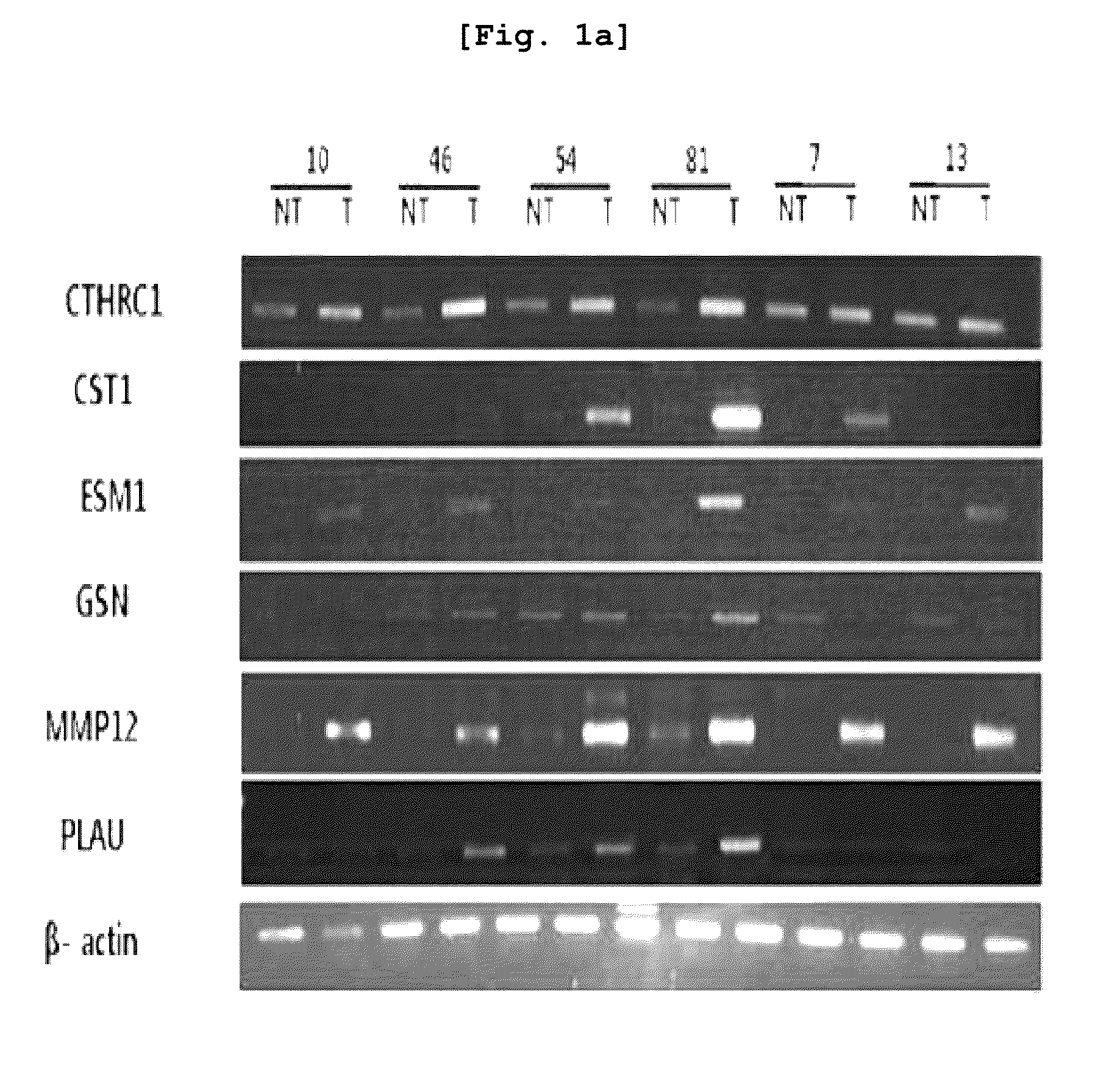
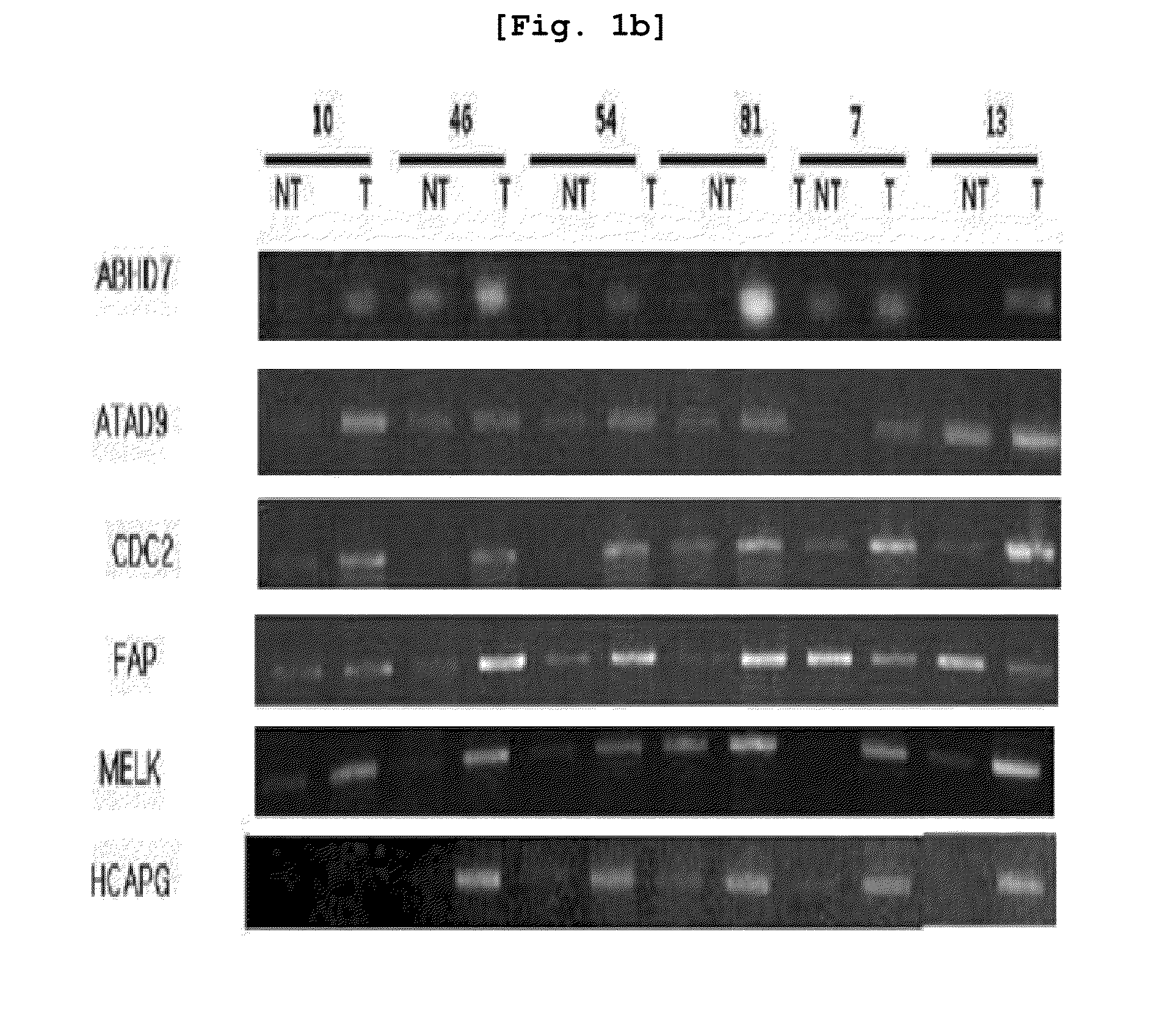
[0112]For cDNA microarray experiment, primary hepatocellular carcinoma (HCC) tissues were obtained from forty liver cancer patients at Seoul St. Mary's Hospital and Chonbuk National University Hospital. The normal liver tissues were obtained from patients other than liver cancer patients at Seoul St. Mary's Hospital. The tissue samples were frozen in liquid nitrogen. The total RNA was isolated using RNeasy mini kit (Qiagen, Hilden, Germany). Differences in gene expression were then investigated based on the cancer tissue of the 40 liver cancer patients and the neighboring tissues using cDNA microarrayer.

example 2

Isolation of mRNA from Tissues and Cells

[0113]For RT-PCR, normal liver cells and liver cancer cells were collected from twenty liver cancer patients and mRNA was isolated from total forty tissues.

[0114]First, upon extracting the tissues with surgical incision, blood was immediately removed in sterilized in PBS, and frozen in liquid nitrogen. After that, the total mRNA was isolated by Guanidinium method and single-step RNA isolation was carried out. The isolated total mRNA was quantified using spectrophotometer, and reserved in −70 C refrigerator for later use.

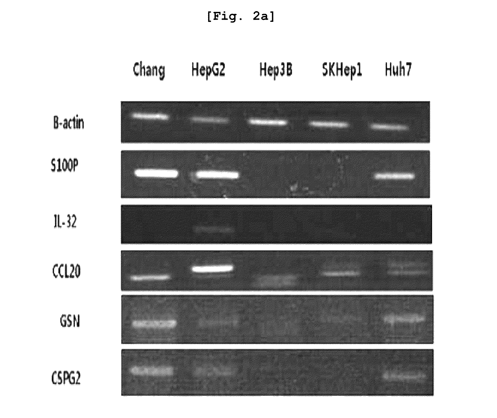
[0115]Total five liver cancer lines were selected (Chang, HepG2, Hep3B, SKHep1, Huh7) and distributed from Korean Cell Line Bank (KCLB) with address at 28, Yeongun-dong, Jongno-gu, Seoul, Republic of Korea.

[0116]10% fetal bovine serum (FBS, Hyclon) and penicillin streptomycin (1 mg / ml, Sigma) were respectively added to optimum culture medium, i.e., DMEM (Invitrogen) or RPMI1640 (Invitrogen), cultured for 5 to 6 days, and total ...

example 3

Comparison of Gene Expression Using RT-PCR

[0117]RT-PCR was carried out regarding the liver cancer-specific over-expression genes which were selected based on the result of Example 1.

[0118]For the purpose of preparing primers, the entire DNA sequences of the respective genes were obtained from core nucleotide (http: / / www.ncbi.nlm.nih.gov.) of NCBI, and the primer sequences of these genes were designed through Prmer3 program. Using the primers designed as explained above, PCR (polymerase chain reaction) was carried out to thereby investigate the levels of expression of the respective genes. The respective primer sequences are illustrated in Table 1 provided above.

[0119]For RT-PCR, cDNA was prepared, from the mRNA extracted from the tissues and cell lines of Example 2 and through reverse transcription (RT) reaction. The cDNA was prepared using cDNA Synthesis kit (AccuAcript High Fidelity 1st Stand cDNA Synthesis Kit, STRATAGENE).

[0120]Using the prepared cDNA and primers, RT-PCR (1 cycl...


PUM


Abstract

The present invention relates to the elucidation of a gene that can act as a novel marker for liver cancer diagnosis and to diagnostic and prognostic measurements of liver cancer using the same. More specifically, it relates to a diagnosis kit that enables diagnostic and prognostic measurement of a liver cancer using a preparation that measures expression levels of at least one gene selected from a group of liver cancer diagnosis markers consisting of S100P, NK4, CCL20, CSPG2, PLAU, MMP12, ESM-1, ABHD7, HCAPG, CXCL-3, Col5A2, MAGEA, GSN, CDC2, CST1, MELK, ATAD2, FAP and MSN and / or a method for diagnostic and prognostic measurement of liver cancer using the same. These have been discovered using normal liver tissues and liver cancer tissues collected from the same liver cancer patient of the present invention and represent the markers whose accuracy and reliability have been greatly improved as markers of liver cancer. The markers of the present invention can be used for the accurate diagnosis and prognosis of liver cancer.

Description

TECHNICAL FIELD[0001]The present invention relates to a biomarker specific to liver cancer and use thereof.BACKGROUND ART[0002]In Southeastern countries including South Korea, Japan, Taiwan, and China, the largest population of patients suffers from liver diseases, such as hepatitis, cirrhosis or liver cancer. The death rate by liver cancer is the fourth-highest around the world by 505,000 patients (WTO, 1997). In South Korea, patients with liver cancer occupy third-largest population of cancer patients by 11.5% (Annual Report of Cancer Statistics in Korea in 2002). A liver cancer has been diagnosed so far based on tissue examination or test with a liver cancer marker such as AFP.[0003]Currently, AFP and PIVKA-II are widely known as the biomarkers for diagnosis, prognosis or evaluation of treatment of liver cancer. However, these markers have yet to meet specificity and sensitivity ideals.[0004]AFP (alpha-fetoprotein) in blood has been widely known for its usefulness in the diagnosi...

Claims


Application Information

Patent Timeline
IPC IPC(8): C40B30/04C40B40/10C12Q1/68C07K16/40G01N33/577C07H21/00C07K16/18C07K16/24C40B40/06G01N33/574
CPCC12Q1/6886C12Q2600/118G01N33/57438G01N33/5023C12Q2600/158A61P35/00C12Q1/6837
Inventor SONG, EUN YOUNGLEE, HEE GUYEOM, YOUNG IIJI, NA YOUNGLEE, JUNG IIKANG, MIN A.KIM, YOUNG JOOKANG, YOON HEEKIM, JAE WHA
Owner KOREA RES INST OF BIOSCI & BIOTECH
Learn More