
Systems and methods for tracking and guiding high intensity focused ultrasound beams

Active Publication Date: 2010-10-28
KONINKLIJKE PHILIPS ELECTRONICS NV
View PDF8 Cites 44 Cited by
  • Summary
  • Abstract
  • Description
  • Claims
  • Application Information

AI Technical Summary

Benefits of technology

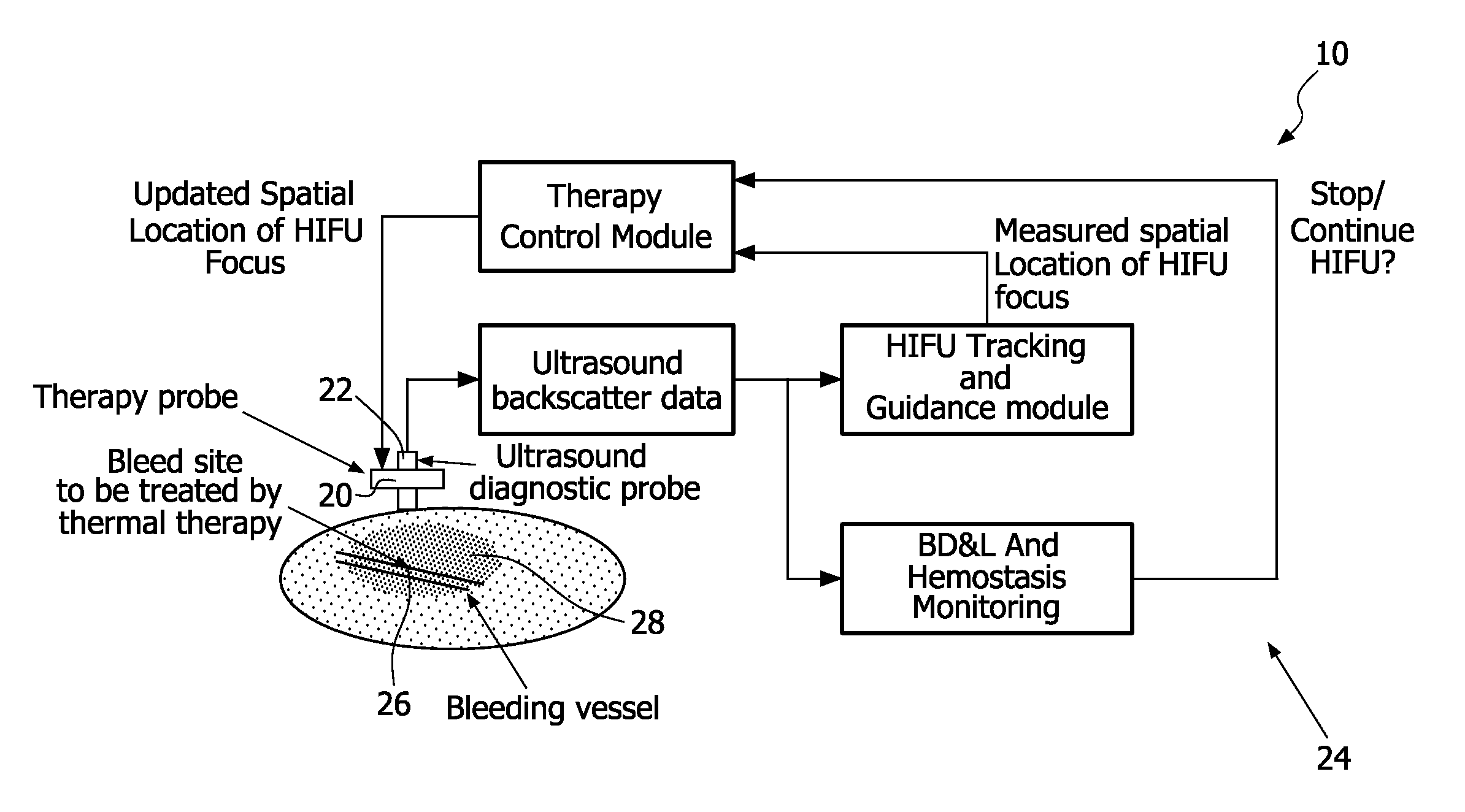
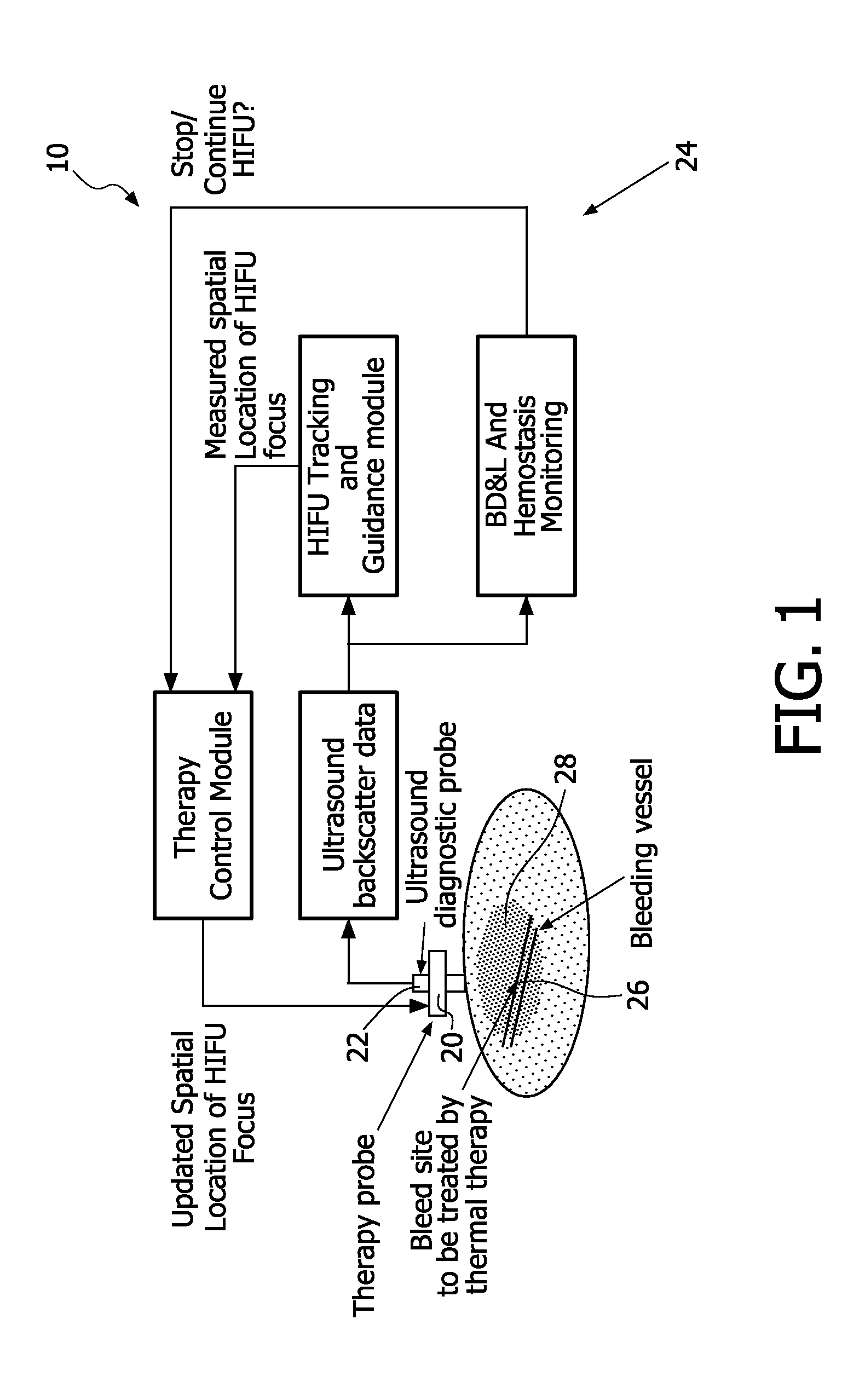
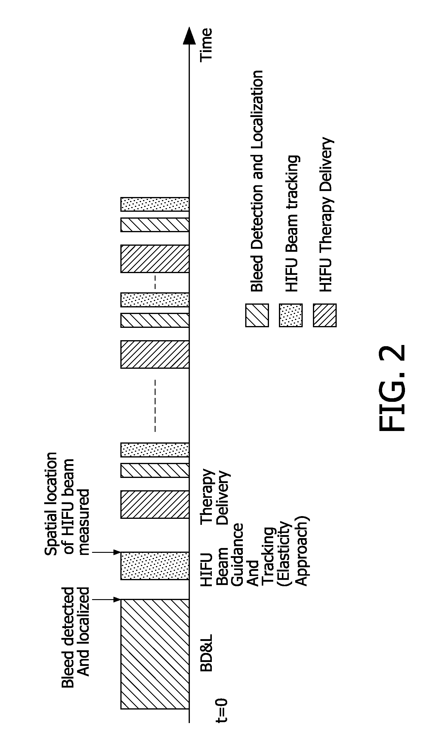
[0017]In general, the systems and methods disclosed, as related to tracking and guiding of an HIFU beam, may be implemented before HIFU therapy commences. Thus, the disclosed systems and methods advantageously facilitate focusing of an HIFU beam relative to a treatment location, while minimizing collateral damage to surrounding tissue. In exemplary embodiments “Bleed Detection and Localization” (BD&L) techniques may be used to detect and localize a desired treatment location. Thus, HIFU beam tracking and beam focusing steps may be cyclically interleaved with BD&L and HIFU beam administration to continuously ensure that HIFU therapy is being delivered to the desired treatment location.

Problems solved by technology

In civilian casualties, exsanguinations due to internal bleeding are the most significant cause of death in trauma victims.
For these emergent care type applications, the device is designed to be operator independent and, therefore, image-based feedback allowing user interaction and control is not available.
Due to tissue heterogeneities and lack of quantitative information on the local thermal and acoustic properties, there exist errors between the intended spatial location of treatment and the actual physical location of the HIFU focus.
Although BD&L techniques may be repeated to determine if a bleed rate has slowed or stopped, these techniques do not provide any quantitative feedback or spatial information relating to the optimal reorientation or refocusing of the therapy relative to the desired treatment location.
However, it has been demonstrated that this information is not always reliable since hyperecho can appear quite some time after the tissue has already been ablated and when the local tissue temperature is close to boiling.
The inhomogeneous structure of tissue would make direct B-mode visualization of the focal region of HIFU preceding boiling even more challenging.
In all cases, the disclosed systems and methods fail to provide adequate means for tracking and guiding therapeutic HIFU beams.
In particular, the noted European publication does not describe a method for identifying the location of HIFU focus to ensure treatment of a desired location.
In addition, the disclosed technique does not detect the HIFU beam focus.
The proposed mechanical frame is specifically applicable for the transducer geometry in obstetrics and gynecology and cannot be extended for other clinical applications of HIFU.

Method used


Image

  • Systems and methods for tracking and guiding high intensity focused ultrasound beams
  • Systems and methods for tracking and guiding high intensity focused ultrasound beams
  • Systems and methods for tracking and guiding high intensity focused ultrasound beams

Examples


Embodiment Construction

)

[0022]Advantageous systems and methods for facilitating High Intensity Focused Ultrasound (HIFU) are provided according to the present disclosure. In general, systems disclosed herein include (i) an HIFU capable transducer, (ii) a diagnostic imaging probe, and (iii) a processor. The disclosed methods typically involve determining the focal position of an HIFU capable transducer and associated tracking / guidance steps / functionalities.

[0023]According to the present disclosure, acoustic radiation force impulse (ARFI) imaging may be used to detect the focal position for an HIFU capable transducer relative to a target area. In exemplary embodiments, the diagnostic imaging probe is used to probe a target area and obtain imaging data before and after inducement of a radiation force relative to the target area. The radiation force is typically induced using the HIFU capable transducer, e.g., using low power sonication. The radiation force causes motion of the target area and the region of g...


PUM


Abstract

The present disclosure provides systems and methods for tracking and guiding high intensity focused ultrasound beams (HIFU). More particularly, the disclosed systems and methods involve use of acoustic radiation force impulse (ARFI) imaging to detect the focal position of an HIFU capable transducer relative to a target area. The focal position may then be 5 compared to a desired treatment location and the orientation and focus of the transducer may be adjusted accordingly so as to reconfigure and / or refocus the HIFU beam relative to the desired treatment location. The desired treatment location may be dynamically determined using bleed detection and localization (BD&L) techniques. Thus, the desired treatment location may be determined using 3D Doppler ultrasound based techniques, wherein changes in quantitative 10 parameters extracted from the Doppler spectra, e.g., Resistance Index (RI), are used to detect and localize a bleeding site for treatment.

Description

Statement of Governmental Interest[0001]The United States government may hold license and / or other rights to the systems and methods claimed herein. Development of the systems and methods claimed herein was supported by DARPA; Funding Opportunity No. DARPA05-01DBAC.BACKGROUND[0002]1. Technical Field[0003]The present disclosure relates to high intensity focused ultrasound (HIFU) applications. More particularly, the present disclosure relates to systems and methods for tracking and guiding HIFU beams. Although exemplary embodiments relate primarily to acoustic hemostasis, the disclosed systems and methods apply to and for any HIFU application, including but not limited to HIFU treatments for uterine fibrosis, cancer, and cardiac arrhythmias, such as atrial fibrillation.[0004]2. Background Art[0005]Blood loss from extremity wounds is the number one cause of preventable battlefield death today. In civilian casualties, exsanguinations due to internal bleeding are the most significant cau...

Claims


Application Information

Patent Timeline
IPC IPC(8): A61N7/00A61B8/00
CPCA61B5/0048A61B8/06A61N7/02A61B8/485A61B2019/5276A61B8/08A61B2090/378
Inventor ANAND, AJAYPETRUZZELLO, JOHN
Owner KONINKLIJKE PHILIPS ELECTRONICS NV
Learn More