Endoscopic digital recording system with removable screen and storage device

Inactive Publication Date: 2010-06-10
ENVISIONIER MEDICAL TECH
View PDF64 Cites 1211 Cited by
  • Summary
  • Abstract
  • Description
  • Claims
  • Application Information

AI Technical Summary

Benefits of technology

[0013]In another embodiment, the present invention provides a portable hand-held endoscopy system adapted for interchangeable use with a variety of endoscopes. The system having an endoscope having a first end and a second end, the first end having an eyepiece and the second end having a viewing end. The system further comprising a battery operated unitary digital camera having an optical input, viewing screen, digital signal processor, memory with embedded software for processing data from the processor and for displaying an image on the viewing screen, and a coupler located at the distal end of the camera for removably connecting to the eyepiece of an endoscope.
[0016]The invention described herein requires only a limited number of components, providing physicians with a portable, versatile, and less expensive system for endoscopic examinations and recording, archiving and sharing the images and videos thereof. Among other things, the present invention eliminates the need for accessory equipment that is infrequently used (e.g., VHS recorders, printers, CD / DVD burners). The system is easily transported to multiple locations, enabling healthcare providers greater versatility in the applications of endoscopic examinations and flexibility of the locations at which they examine patients. In addition to cost savings and flexibility, the high speed data transfer technology facilitates higher speed, lower cost data translation and manipulation, enhancing and expanding the quality of visual documentation generated without the need for special or costly computer systems.
[0019]In a preferred embodiment, the portable LCD device of the present invention (sometimes referred to as “the eGo”) is a mass storage device with full multimedia playback that can be mounted on flexible scopes or other mounts to encourage natural line of site aligned with the viewing field. On board data management and integration with EMRs eliminates frustrating and cumbersome tasks all too often associated with endoscopic data management.
[0020]The portable device may include a viewing screen to permit a user to view or replay images in the form of, for example, still shots or videos, on the portable device. The viewing screen may also be a touch screen to allow for a user to operate the portable device or input information and data into the portable device. For example, using the portable device LCD touch screen allows the user to tag an endoscopic exam to the patient's information without redundant effort. A gravity detecting system may also be used with the portable device to align the still shots and videos during playback and lighting control is optimized for any environment.
[0021]LED lighting technology is provided for brighter lighting capability than in many tabletop Halogen or Metal Halide light sources. Combining the present invention with high powered LED light source provides a convenient, versatile, and scalable endoscopic imaging system for use in multiple exam rooms in an outpatient clinical setting. This combination is also perfect for academic teaching environments and remote endoscopic diagnosis.

Problems solved by technology

These systems are commonly used in doctor's offices, emergency rooms, hospital rooms, and operating rooms, but are very expensive, not easily adapted, and not configured to be easily transported between and among multiple locations.
The cameras currently available to medical professionals are not easily configured for endoscopic imaging.
The camera control unit and accompanying computer and viewing screen are bulky, heavy, and not easily transported to different locations.
In addition to the size and transport limitations, the systems currently available can range in cost from $10,000 or more for just the camera and camera control unit.
In addition to the cost of the camera and camera control unit, the endoscope, and typically a light source must be purchased.
These alternatives, however, limit editing of the images and are not very dynamic.
These systems provide better data manipulation, but can cost more than $20,000, and thus not affordable for a small or cost-limited practice.
Although these systems are classified as portable, they are heavy, cumbersome, and expensive.
Additionally, this aforementioned patent is not readily adapted to the standard fittings of the eyepiece of endoscopes used in medical practices.
While analog devices recorded images, their use came with several limitations including, but not limited to, the inability to easily incorporate into electronic media for sharing and reviewing (like e-mail, electronic medical records, and PowerPoint®) the difficulty in cataloging analog images.
However, these digital systems are extremely bulky and are part of a larger component cart system not allowing for portability or use with multiple devices.
Further, current digital systems do not allow for easy incorporation of electronic medical record data into the device to streamline data management.

Method used


Image

  • Endoscopic digital recording system with removable screen and storage device
  • Endoscopic digital recording system with removable screen and storage device
  • Endoscopic digital recording system with removable screen and storage device

Examples


first embodiment

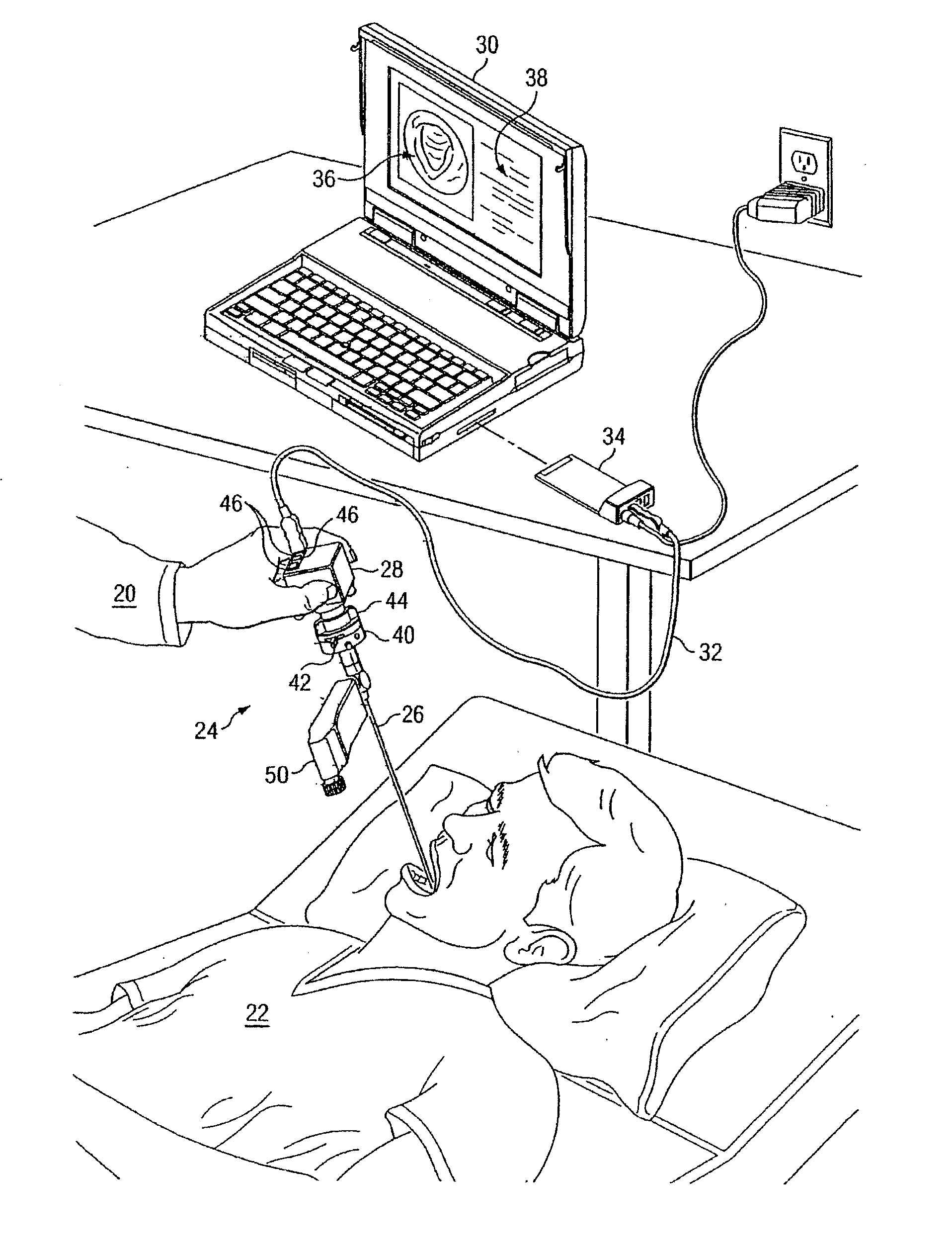
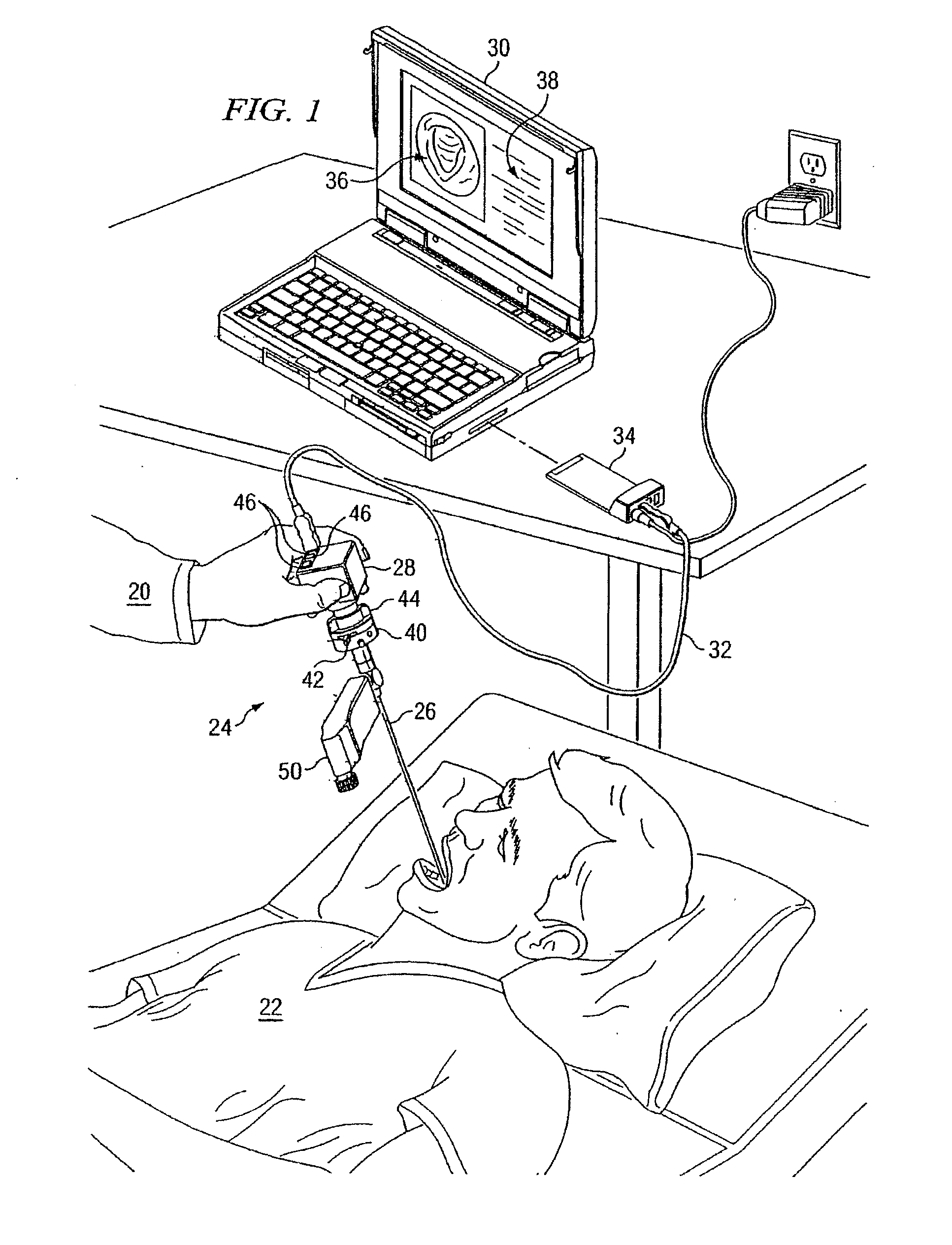
[0067]Referring to the Drawings, and particularly to FIGS. 1 and 2 thereof, there is shown an endoscopic imaging system comprising the invention. Referring specifically to FIG. 1, a physician 20 is shown performing an endoscopic examination of a patient 22 using an endoscopic imaging system 24. An endoscope 26 is inserted into the patient 22. The images seen by the endoscope 26 are received into a portable endoscopic digital camera 28 capable of high speed data transfer and then transmitted to a computer 30 by means of a high speed data transfer connection cable 32, for example a USB cable. The cable 32 connects into a multifunctional interface card 34, which also supplies power to the camera 28. Alternately, the high speed data transfer connection cable can connect directly to a computer or similar computing device without a multifunctional interface card if said high speed data transfer connection is built into the computing device. Examples of the high speed data transfer consist...

second embodiment

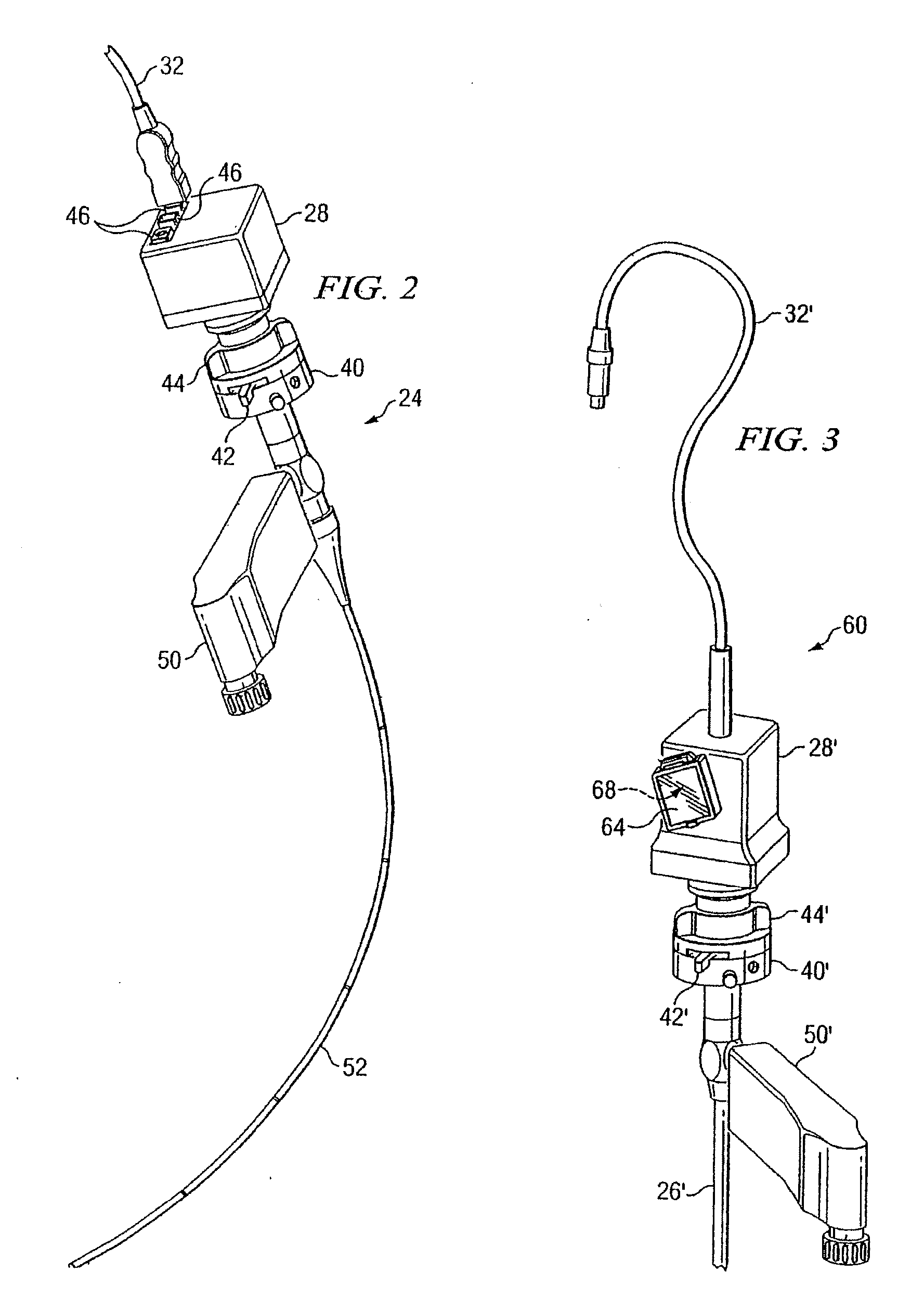
[0070]Referring now to FIG. 3, there is shown an endoscopic imaging system 60 comprising the invention. Many of the component parts of the endoscopic imaging system 60 are substantially identical in construction and function to component parts of the endoscopic imaging system 24 illustrated in FIGS. 1 and 2 and described hereinabove in conjunction therewith. Such identical component parts are designated in FIG. 3 with the same reference numerals utilized above in the description of the endoscopic imaging system 24, but are differentiated there from by means of a prime (′) designation.

[0071]The endoscopic imaging system 60 differs from the endoscopic imaging system 24 of FIGS. 1 and 2 in that the endoscopic imaging system 60 includes a camera 28′ with an on-board LCD screen 64 and on-board one-touch camera controls with embedded software for manipulating, enhancing, and adjusting the data. It will be appreciated that the screen may be an LCD screen, LED screen or any other similar mo...


PUM


Abstract

A medical imaging recording and viewing system adapted for use interchangably with a variety of endoscopes. The system includes a portable hand-held device that may be removably placed within a cradle affixed to the endoscope. An interface between an image acquisition device of the endoscope and the portable device permits images to be transmitted to and stored within the portable device. The portable device also may include a viewing screen to permit a user to view the images. A docking station may receive the portable device for transferring images to a separate viewing device and for recharging the battery of the portable device.

Description

BACKGROUND OF THE INVENTION[0001]This application claims the benefit of U.S. Provisional Application No. 61 / 248,443, filed Oct. 3, 2009, and is a continuation in part of U.S. Design Patent Application No. 29 / 322,352, filed Aug. 1, 2008, U.S. Design Patent Application No. 29 / 322,349, filed Aug. 1, 2008, U.S. Design Patent Application No. 29 / 322,353, filed Aug. 1, 2008, and U.S. patent application Ser. No. 11 / 768,965, filed Jun. 27, 2007 which is a continuation-in-part of International Patent Application No. PCT / US2005 / 047281, filed Dec. 28, 2005 and which claims the benefit of U.S. Provisional Application No. 60 / 639,451, filed Dec. 28, 2004, the entirety of all of these applications are hereby incorporated by reference.FIELD OF THE INVENTION[0002]This invention relates generally to portable, hand-held endoscopic recording systems, and more particularly to a digital endoscopic recording system having a docking station with a removable LCD screen with internal memory for storage and re...

Claims


Application Information

Patent Timeline
IPC IPC(8): A61B1/04
CPCA61B1/00052A61B1/042A61B1/00105
Inventor MELDER, PATRICK C.
Owner ENVISIONIER MEDICAL TECH
Why Eureka
Try Eureka