Expansion and Differentiation of Mesenchymal Stem Cells
a mesenchymal stem cell and expansion and differentiation technology, applied in the field of expansion and differentiation of mesenchymal stem cells, can solve the problems of limited amount of msc that can be retrieved, limited amount of msc, and limitation of msc surface molecules seen in laboratory, and achieve the effect of high capacity to form cartilag
- Summary
- Abstract
- Description
- Claims
- Application Information
AI Technical Summary
Problems solved by technology
Method used
Image
Examples
example 1
Isolation of Mesenchymal Stem Cells from Human Bone Marrow
Objective
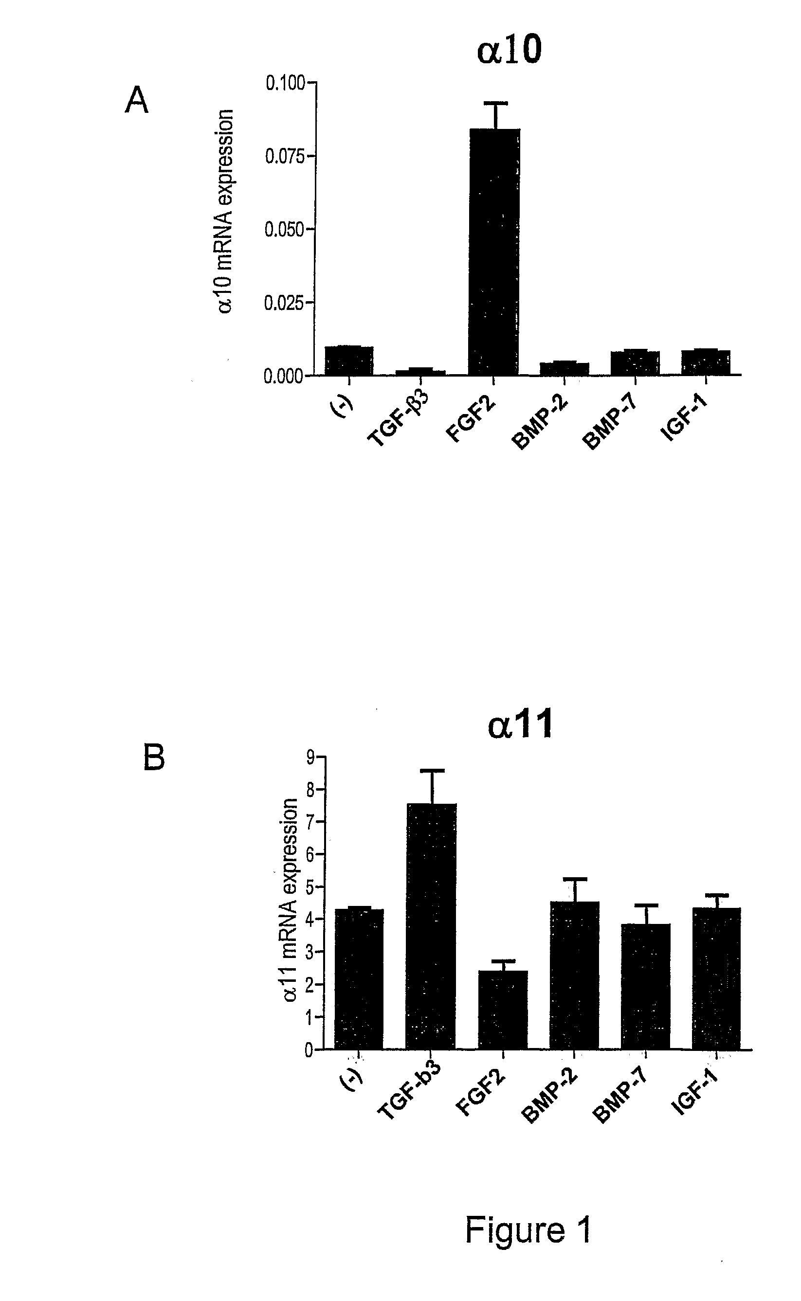
[0266]The objective with this example was to demonstrate that certain growth factors could affect the mRNA expression of integrin alpha10 on human MSC.
Materials and Methods
[0267]A. Isolation of Mesenchymal Stem Cells from Human Bone Marrow
[0268]Posterior iliac aspirations were performed on healthy volunteers for adult bone marrow (BM) collection. The human bone marrow cells were diluted in equal amount of PBS (with Ca2+ and Mg2+) (GibcoBRL, Paisley, UK), 0.6% NaCitrate (Sigma, Sweden), 0.1% BSA (SERVA Electrophoresis GmbH, Heidelberg, Germany) and 100 U / ml DNase (Sigma, Sweden).
[0269]Mononuclear cells (MNCs) were isolated by layering the bone marrow cells on a density gradient (Lymphoprep™, density 1.077 g / ml, Nycomed, Norway) accordingly to the manufactures descriptions.
[0270]MNC were washed twice in PBS and resuspended in MEM α-Medium (GibcoBRL, Paisley, UK) with 20% FCS, 100 U / ml Penicillin and 100 μg / ml streptomy...
example 2
Regulation of Alpha 10 and Alpha 11
Objective
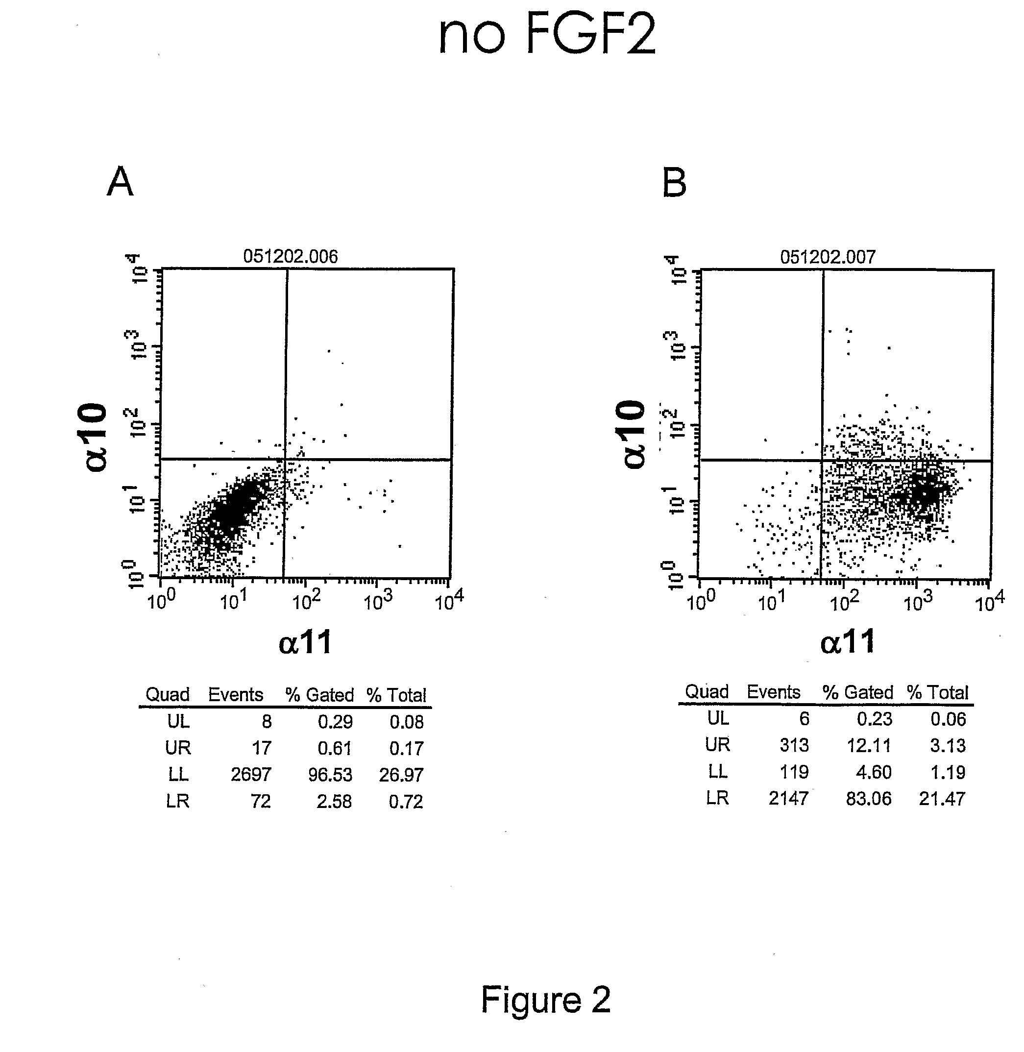
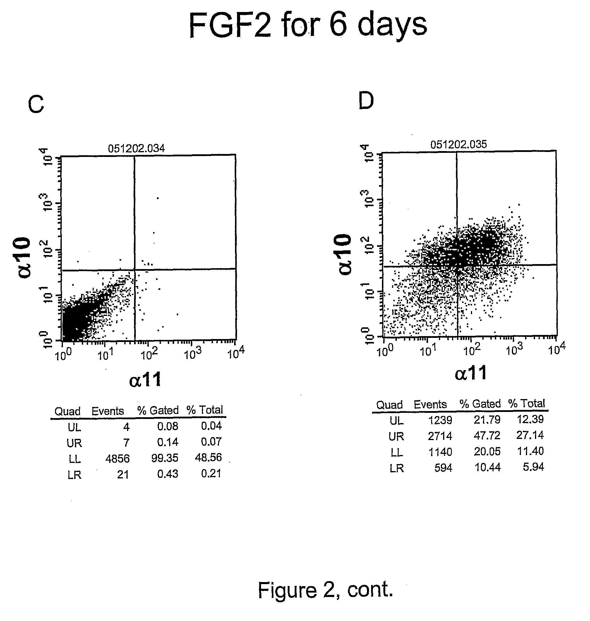
[0276]The objective with this example was to demonstrate that FGF2 regulates the cell-surface expression of integrin alpha10 and alpha11 in hMSC
Materials and Methods
[0277]Human MSCs were isolated and cultured as described in Example 1. The primary antibodies used were mAb365 mIgG2a (anti-alpha 10), with isotype control IgG2a, C09-biotin (anti-alpha11) and the isotype control CT17-biotin at a concentration of 1 μg / ml (both antibodies from Bioinvent Int AB, Sweden).
[0278]Secondary antibodies used were Cy™5 conjugated anti-mIgG (Jackson ImmunoResearch, Pennsylvania) and PE conjugated Streptavidin (BD, San Jose, Calif.). The FACS staining was done according to the manufacturer's instructions. The cell marker expression was detected with a FACSort (13D, San Jose, Calif.) and analyzed using the CellQuest® software (BD, San Jose, Calif.).
Results
[0279]FGF2 treatment of hMSC for 6 days resulted in an increase from 12% to 70% of alpha10 positive cel...
example 3
Human MSC with an Enhanced Chondrocyte Potential
Objective
[0281]The objective with this example was to demonstrate that integrin alpha10-high / alpha11-low hMCSs has an enhanced chondrogenic potential compared to integrin alpha10-low / alpha11-high hMCSs
Materials and Methods
[0282]Human MSCs were isolated as described in example 1. At day 7 after isolation MSCs were cultured in presence or absence of 10 ng / ml FGF2 (BioSource Europe SA, Belgium) for 14 days. The cells were stained and FACS-analyzed as described in example 2.
[0283]MSCs were induced to chondrogenic phenotype in pellet mass culture using 2×10⁵ cells / pellet in DMEM (GibcoBRL, Paisley, UK) supplemented with 1× Insulin-transferrin sodium selenite (Sigma, Sweden), 0.1 μM dexamethasone (Sigma, Sweden), 50 μM ascorbic acid (Sigma, Sweden), 1 mg / ml Linoleic acid-bovine serum albumin (Sigma, Sweden), 1% Nonessential AA (GibcoBRL, Paisley, UK), 100 U / ml Penicillin, 100 μg / ml Streptomycin (GibcoBRL, Paisley, UK) and 10 ng / ml TGF-β3 (R&...
PUM
| Property | Measurement | Unit |
|---|---|---|
| density | aaaaa | aaaaa |
| detection threshold | aaaaa | aaaaa |
| density | aaaaa | aaaaa |
Abstract
Description
Claims
Application Information
 - R&D
- Intellectual Property
- Life Sciences
- Materials
- Tech Scout
- Unparalleled Data Quality
- Higher Quality Content
- 60% Fewer Hallucinations
Browse by: Latest US Patents, China's latest patents, Technical Efficacy Thesaurus, Application Domain, Technology Topic, Popular Technical Reports.
© 2025 PatSnap. All rights reserved.Legal|Privacy policy|Modern Slavery Act Transparency Statement|Sitemap|About US| Contact US: help@patsnap.com


