Devices and processes for nucleic acid extraction
a nucleic acid and process technology, applied in the direction of positive displacement liquid engine, sugar derivative preparation, microorganism lysis, etc., can solve the problems of high cost, time-consuming manual methods, and significant risk of sample contamination
- Summary
- Abstract
- Description
- Claims
- Application Information
AI Technical Summary
Benefits of technology
Problems solved by technology
Method used
Image
Examples
example 1
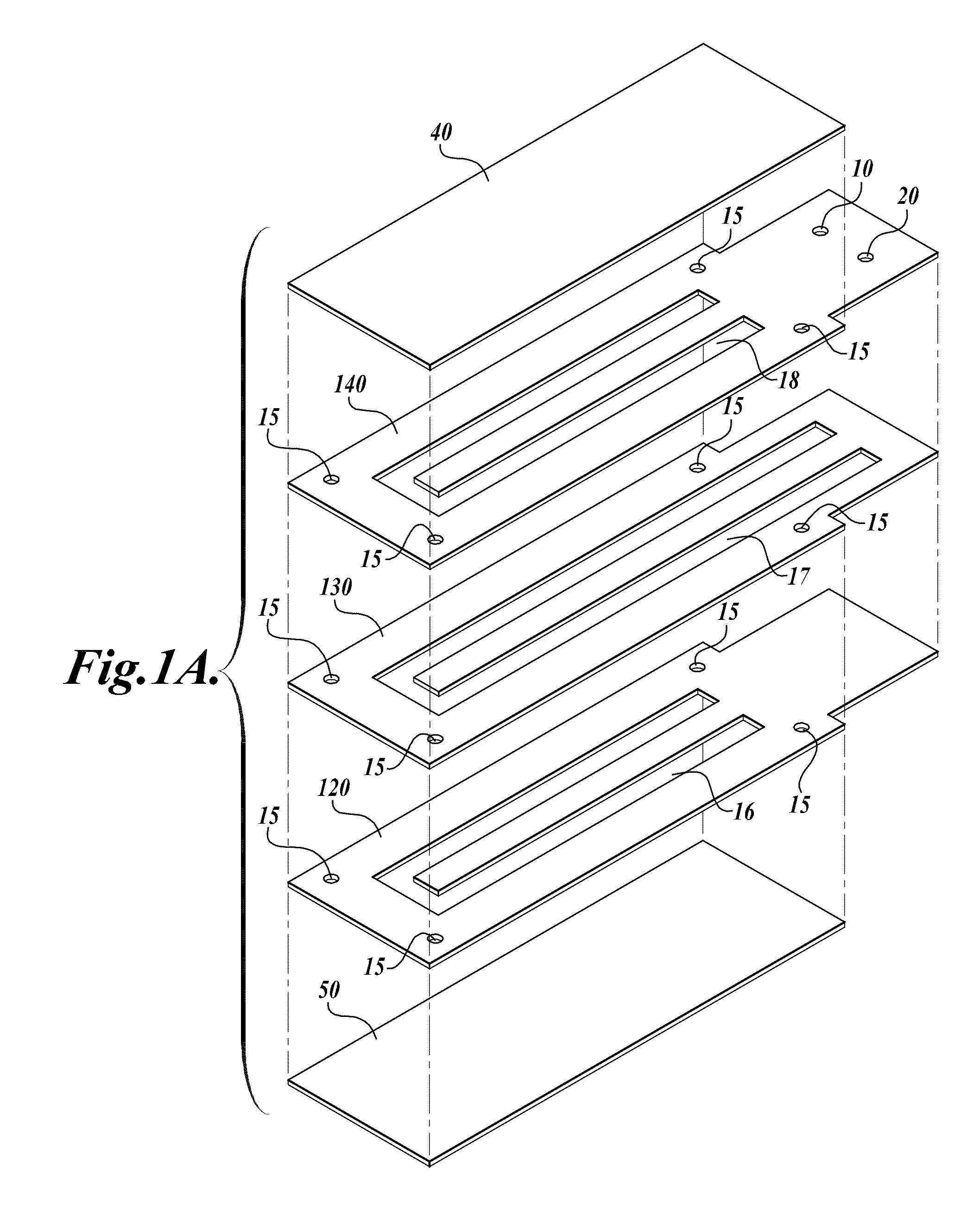
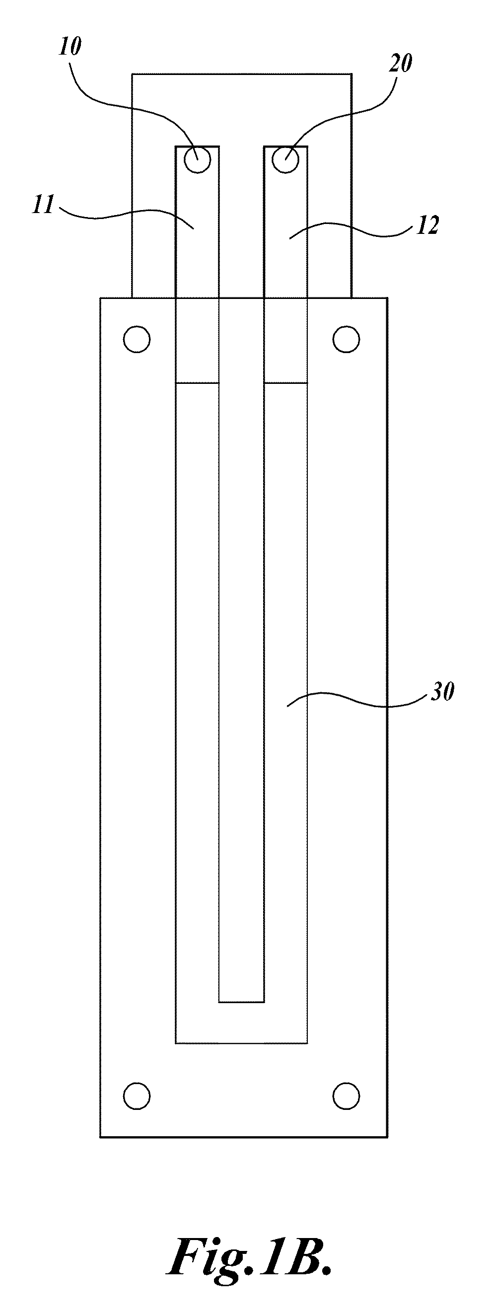
[0113]A compression-sealed device was constructed as shown in FIGS. 3A and 3B. A silicone rubber block was die cut to create a serpentine channel (S-channel) that fit within the area of a standard glass slide. The channel had an overall footprint width of 25.3 mm and length of 75.5 mm. The device was assembled with glass microscope slides on both sides of the S-channel. Acrylic U-Channel was used to provide sufficient clamping pressure to prevent leaks between the glass and the silicone rubber. The two glass slides were separated by a distance of 62.5 mils (1587.5 μm), resulting in an S-channel volume (binding chamber volume)=1444 μL. The area covered by the S-channel was 910 mm2. With two glass surfaces, the total glass area exposed to liquids=1820 mm2, which is approximately equivalent to the area of one surface of a single glass slide (1910 mm2). The exterior dimensions of the device without added fittings were approximately 76 mm by 30 mm by 10 mm (thickness).
[0114]Fluid is port...
example 2
[0115]Twenty μL Subtilisin protease (10 mg / mL stock; obtained from Sigma-Aldrich) is mixed with 200 μL whole blood. 200 μL lysis reagent (6M guanidine hydrochloride, 50 mM citric acid pH 6.0, 20 mM EDTA, 10% Tween-20, 3% TRITON X-100) is added. The solution is mixed well using a pipettor, incubated at room temperature for 15 minutes, and 200 μL pure ethanol is added. The contents of the tube are mixed well.
[0116]Using a pipette, the entire sample is slowly loaded in to extraction device through one port. The sample is allowed to remain in contact with the glass surfaces for at least 1 minute and up to 20 minutes (most binding occurs in the first minute). The sample then is removed from the device. The binding chamber is filled with wash buffer 1 (lysis buffer without detergents diluted with equal volumes of water and 100% ethanol). The buffer is removed, and the wash is repeated two more times for a total of 3 washes. The binding chamber is then filled with wash buffer 2 (20 mM Tris...
example 3
[0119]DNA extraction from whole blood and platelet-rich plasma were compared using glass slides (Nanassy et al., Anal. Biochem. 365:240-245, 2007) and a commercially available spin column kit comprising a glass fiber binding matrix mounted in a small column that fits in a microfuge tube (obtained from Qiagen). Commercially available buffers (Qiagen) were used in most of these studies except as noted for the platelet-rich plasma samples. Platelet rich plasma was prepared from whole blood by the Puget Sound Blood Center (PSBC) according to routine blood center protocols, and the whole blood was also drawn at the PSBC.
[0120]To isolate DNA on glass slides, 200-μL samples of whole blood or platelet-rich plasma were mixed with 400 μg of Proteinase K and 200 μL Buffer AL (which contains guanidine hydrochloride) and incubated at 55° C. for 15 minutes. After the incubation, 200 μl of 100% ethanol was added resulting in a total lysate volume of 600 μl. For some experiments the proportions of ...
PUM
| Property | Measurement | Unit |
|---|---|---|
| Temperature | aaaaa | aaaaa |
| Temperature | aaaaa | aaaaa |
| Volume | aaaaa | aaaaa |
Abstract
Description
Claims
Application Information
 - R&D
- Intellectual Property
- Life Sciences
- Materials
- Tech Scout
- Unparalleled Data Quality
- Higher Quality Content
- 60% Fewer Hallucinations
Browse by: Latest US Patents, China's latest patents, Technical Efficacy Thesaurus, Application Domain, Technology Topic, Popular Technical Reports.
© 2025 PatSnap. All rights reserved.Legal|Privacy policy|Modern Slavery Act Transparency Statement|Sitemap|About US| Contact US: help@patsnap.com


