Diagnosis and prognosis of cancer based on telomere length as measured on cytological specimens
a telomere length and cytology technology, applied in the field of cell biology, molecular biology, and cancer prognosis and diagnosis, can solve the problem of difficult diagnosis of lung cancer by cytology alone, and achieve the effect of longer telomere length
- Summary
- Abstract
- Description
- Claims
- Application Information
AI Technical Summary
Benefits of technology
Problems solved by technology
Method used
Image
Examples
example 1
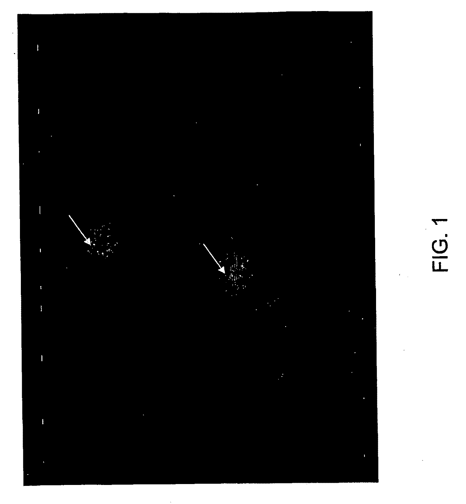
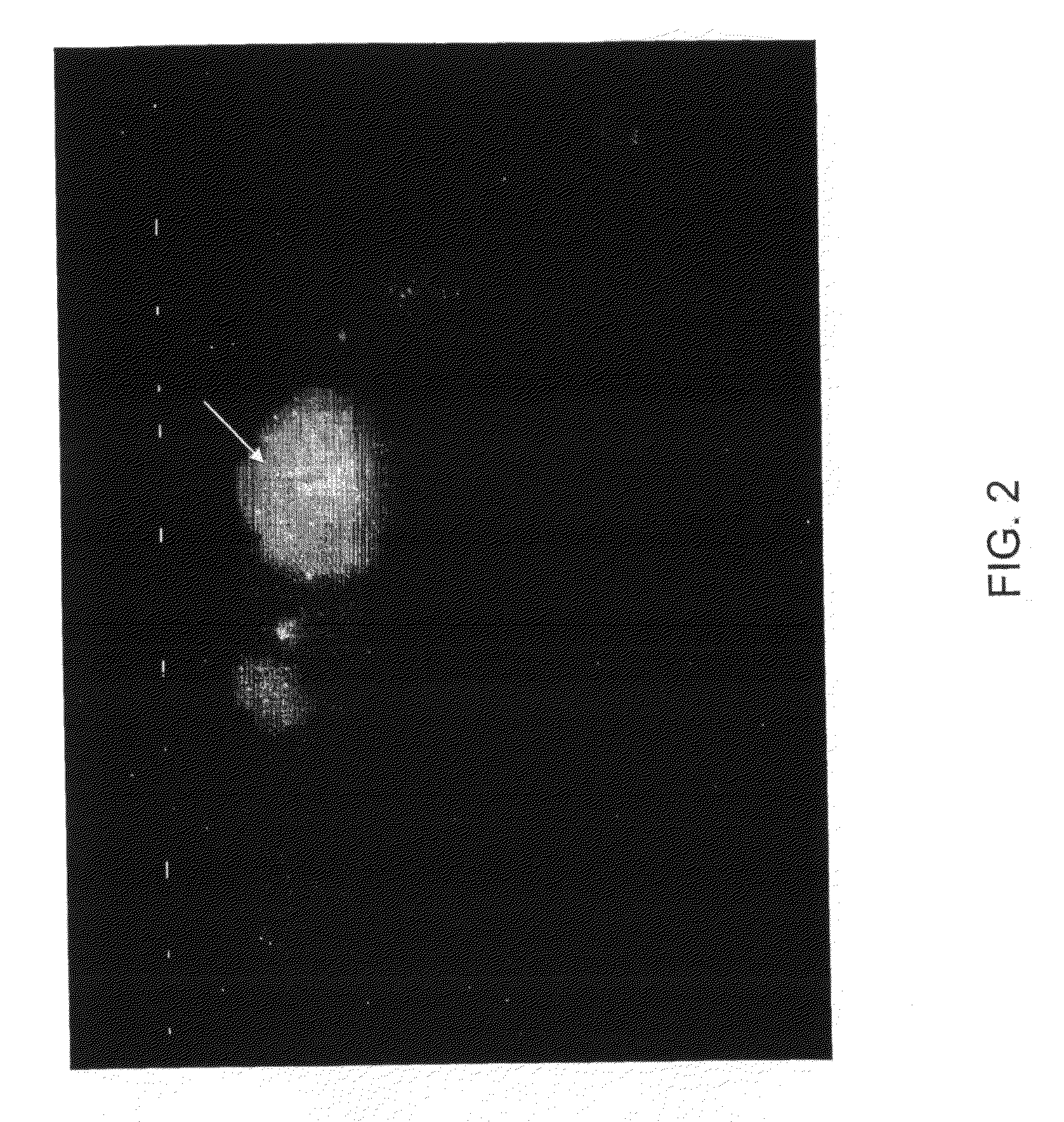
Fluorescence In Situ Hybridization for Telomeres
[0158]Slides were pretreated in 2× sodium saline citrate (SSC) for 2 minutes at 73° C. Slides were then digested with 0.5 mg / ml protease (Vysis Inc., Downers Grove Ill.) in 1×PBS, pH 2.0 at 37° C. for 8 minutes, washed with water and rinsed in 1×PBS for 5 minutes, fixed in 1% formaldehyde in 1×PBS and again rinsed in 1×PBS for 5 minutes. Slides were then denatured with 70% formamide in 2×SSC at 74° C. for 5 minutes and quenched with cold 70% ethanol for 2 minutes, then dehydrated and air-dried. The PNA telomere probe mixture (Applied Biosystems, MA) was denatured at 74° C. for 5 minutes, applied to denatured slide, coverslipped, sealed with rubber cement and incubated in a humid chamber at room temperature for hybridization. After hybridization for 4 hours, slides were washed at 57° C. in 0.1% Tween 20 in 1×PBS for 30 minutes and then rinsed in 0.1% Tween 20 in 2×SSC for 1 minute at room temperature, and air-dried. Slides were then cou...
example 2
Image Capturing
[0159]The images are captured by fluorescent microscope (Leica DMLB) equipped with 100-watt mercury lamp and Vysis filter set DAPI single band pass (DAPI counterstain), and Green single band pass at 63×. Ten (urine) or fifty (bronchial) non-overlapping cells and nuclei with distinct signals were captured using the same exposure, gain and offset (Exposure=1 second, Gain=50, Offset=0). These images were then converted into 8-bit grey scale TIFF files and are then analyzed with Metamorph Offline Software (Universal Imaging Corporation, PA). The software measured the average integrated intensity of total nuclear telomeres as a measure of telomere length.
example 3
Telomere Length in Bladder Cancer
[0160]Telomere length was assessed in individuals known to have bladder cancer, suspected of having bladder cancer, or not having bladder cancer. Table 3 provides data concerning the measurements for the average integrated intensity (the area of the nucleus) over the average area (the intensity of the telomeres), which determines the average intensity of TL.
[0161]The column entitled “Abnormal Cells” refers to the number of cells that were considered abnormal by UroVysion FISH analysis standards (wherein a cell is classified as abnormal when the ploidy is greater or less than diploid in two or more chromosomes or 9p21. It is noteworthy that the individuals with patient ID numbers 9, 10, and 11, for example, were classified as normal by UroVysion FISH, whereas the methods of the present invention classified them as having cancer (based on average intensities of 4.5, 4.55, and 4.97, respectively), and upon follow-up two of three individuals were diagnos...
PUM
| Property | Measurement | Unit |
|---|---|---|
| volume | aaaaa | aaaaa |
| volume | aaaaa | aaaaa |
| volume | aaaaa | aaaaa |
Abstract
Description
Claims
Application Information
 - R&D
- Intellectual Property
- Life Sciences
- Materials
- Tech Scout
- Unparalleled Data Quality
- Higher Quality Content
- 60% Fewer Hallucinations
Browse by: Latest US Patents, China's latest patents, Technical Efficacy Thesaurus, Application Domain, Technology Topic, Popular Technical Reports.
© 2025 PatSnap. All rights reserved.Legal|Privacy policy|Modern Slavery Act Transparency Statement|Sitemap|About US| Contact US: help@patsnap.com


