Compositions and Method for Brain Specific Targeted Delivery of Therapeutic Agents
a therapeutic agent and brain technology, applied in the direction of drug compositions, biocide, genetic material ingredients, etc., can solve the problems of inflammatory reactions, unfavorable delivery of therapeutic agents, and undesirable viral vectors
- Summary
- Abstract
- Description
- Claims
- Application Information
AI Technical Summary
Problems solved by technology
Method used
Image
Examples
example 1
Large-Scale Manufacture of Polynucleotides Encoding a Green Fluorescent Protein (GFP)
[0047]Plasmid DNA (pEGFP-N1; from Clontech, GenBank Accession #U55762) was isolated from bacteria using the EndoFree Qiagen plasmid maxi purification kit following the manufacturers protocol (from Qiagen). pEGFP-N1 includes a polynucleotide encoding EGFP under the control of CMV, a constitutive promoter. To produce larger quantities bacteria comprising the pEGFP-N1 plasmid were grown in 100 mL LB batches.
example 2
Nanoparticle Fabrication
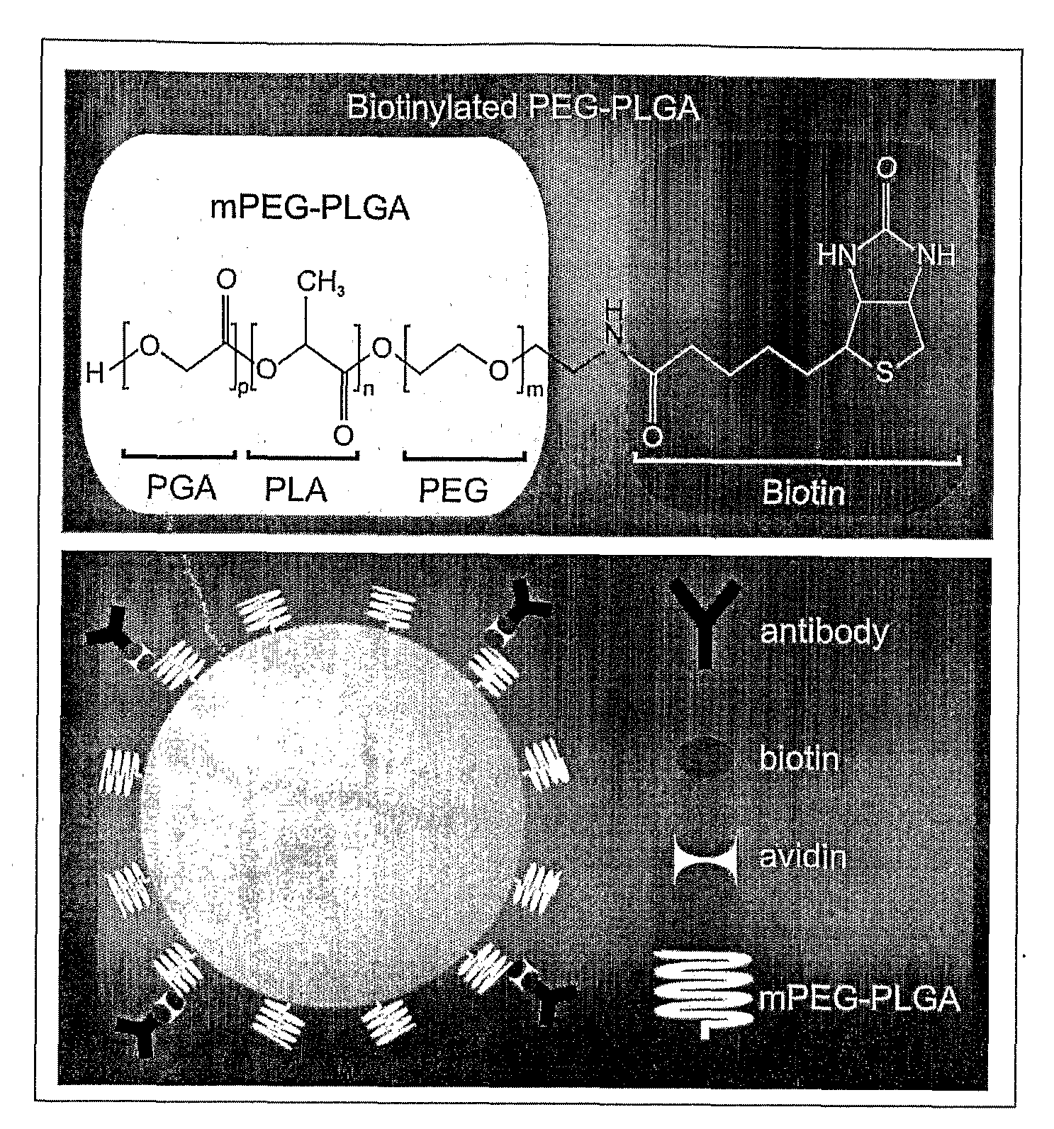
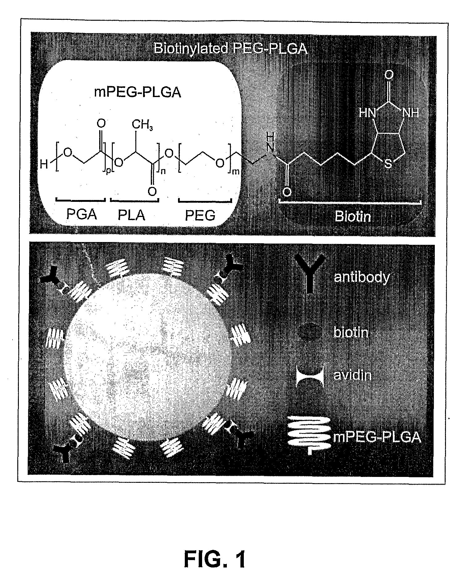
[0048]PLGA and mPEG-PLGA nanoparticles of uniform size (i.e., 100 nm, 150 nm and 250 nm) were synthesized. PLGA nanospheres (of approximately 250 nm diameter) were produced as follows. 4 mg acetylated bovine serum albumin (BSA) (Sigma) was dissolved in 66.7 μL of water. 400 μg DNA (pEGFP-N1) was dissolved in 40 μL TE 7.4 buffer. 12 mg of PLGA (MW: 30,000; 50:50 (from Boehringer Ingelheim)) polymer was dissolved in 400 μL chloroform. If nanoparticles capable of carrying specific surface monoclonal antibodies (mAbs) were desired, 1-2% of the starting mPEG-PLGA (or PLGA) polymer was replaced by biotin-PEG-PLGA (or biotin-PLGA). The DNA and BSA were added together to the dissolved PLGA with a glass Pasteur pipette while the PLGA was sonicated. The emulsion was sonicated for 30 seconds at 45-50 watts in an ice bath. The primary water / oil (w / o) emulsion was added to 3.33 mL of 2.5% polyvinyl alcohol (PVA); MW: 50,000) with a glass Pasteur pipette while the PVA was ...
example 3
Characterization of Nanoparticles
[0053]Nanospheres were examined using scanning electron microscopy (SEM) to illustrate the size of nanospheres. Samples were prepared by dropping an aliquot of the nanosphere suspensions of Example 1 onto a polished aluminum stub. Excess solution was wicked off with filter paper leaving a thin coating of nanospheres. A thin layer of a gold coating covering the particles was applied by sputtering gold at 10 mA and 6 V (Model Huml; Technics) for 0.5 to 6 minutes. The surface morphology of the nanospheres was examined by High Resolution SEM (Hitachi S-4700-II) at 5-15 kV. The particles ranged in size from about 200 nm to about 250 nm, appeared round and showed little evidence of aggregation.
[0054]Particle sizing, particle distribution, and zeta potential measurements were performed using a Brookhaven ZetaPlus Analyzer. Nanoparticle size standards from Duke Scientific were prepared according to the certification records and used to check the performance ...
PUM
| Property | Measurement | Unit |
|---|---|---|
| diameter | aaaaa | aaaaa |
| diameter | aaaaa | aaaaa |
| diameter | aaaaa | aaaaa |
Abstract
Description
Claims
Application Information
 - R&D
- Intellectual Property
- Life Sciences
- Materials
- Tech Scout
- Unparalleled Data Quality
- Higher Quality Content
- 60% Fewer Hallucinations
Browse by: Latest US Patents, China's latest patents, Technical Efficacy Thesaurus, Application Domain, Technology Topic, Popular Technical Reports.
© 2025 PatSnap. All rights reserved.Legal|Privacy policy|Modern Slavery Act Transparency Statement|Sitemap|About US| Contact US: help@patsnap.com


