Methods of diagnosing synovial disease in a mammal by detecting bacterial DNA in synovial tissues from dogs with inflammatory knee arthritis and degenerative anterior cruciate ligament rupture

a technology of synovial tissue and mammalian cells, which is applied in the field of human-made kits and methods for detecting synovial disease in the knee of a human, can solve the problems of complex interaction between bacteria and the host immune system, unrecognized specific mechanism involving bacterial triggering of joint inflammation, and inability to detect bacterial dna in synovial tissues,

Inactive Publication Date: 2008-02-21
WISCONSIN ALUMNI RES FOUND
View PDF5 Cites 9 Cited by
  • Summary
  • Abstract
  • Description
  • Claims
  • Application Information

AI Technical Summary

Benefits of technology

[0040] One aspect of the invention is a method of diagnosing persistent, chronic synovitis in the joint of a mammal comprising the steps or acts of providing a test sample comprising synovial fluid, cell or tissue from the joint of a mammal; quantifying the concentration of one or more protein or mRNA biomarkers being cathepsin K, MMP-2, MMP-9, cathepsin S, TRAP, invariant chain, CD4+ T-lymphocytes, CD8+ T-lymphocytes, CD44+ mo...

Problems solved by technology

However, any specific mechanism involving bacterial triggering of joint inflammation is not well understood.
In a proportion of patients infected with Lyme disease, persistent and severe synovitis develops that is refractory to known treatments.
Interaction between bacteria and the host immune system is complex.
However, it is currently unclear what specific immune mechanism perpetuates joint inflammation in all forms of chronic inflammatory arthritis.
However, synovitis in the contralateral stifle remains unexplained.
Critical evaluation of those results may not be available in abundance.
Apparently, such investigations have not been performed in d...

Method used


Image

  • Methods of diagnosing synovial disease in a mammal by detecting bacterial DNA in synovial tissues from dogs with inflammatory knee arthritis and degenerative anterior cruciate ligament rupture
  • Methods of diagnosing synovial disease in a mammal by detecting bacterial DNA in synovial tissues from dogs with inflammatory knee arthritis and degenerative anterior cruciate ligament rupture
  • Methods of diagnosing synovial disease in a mammal by detecting bacterial DNA in synovial tissues from dogs with inflammatory knee arthritis and degenerative anterior cruciate ligament rupture

Examples


example 1

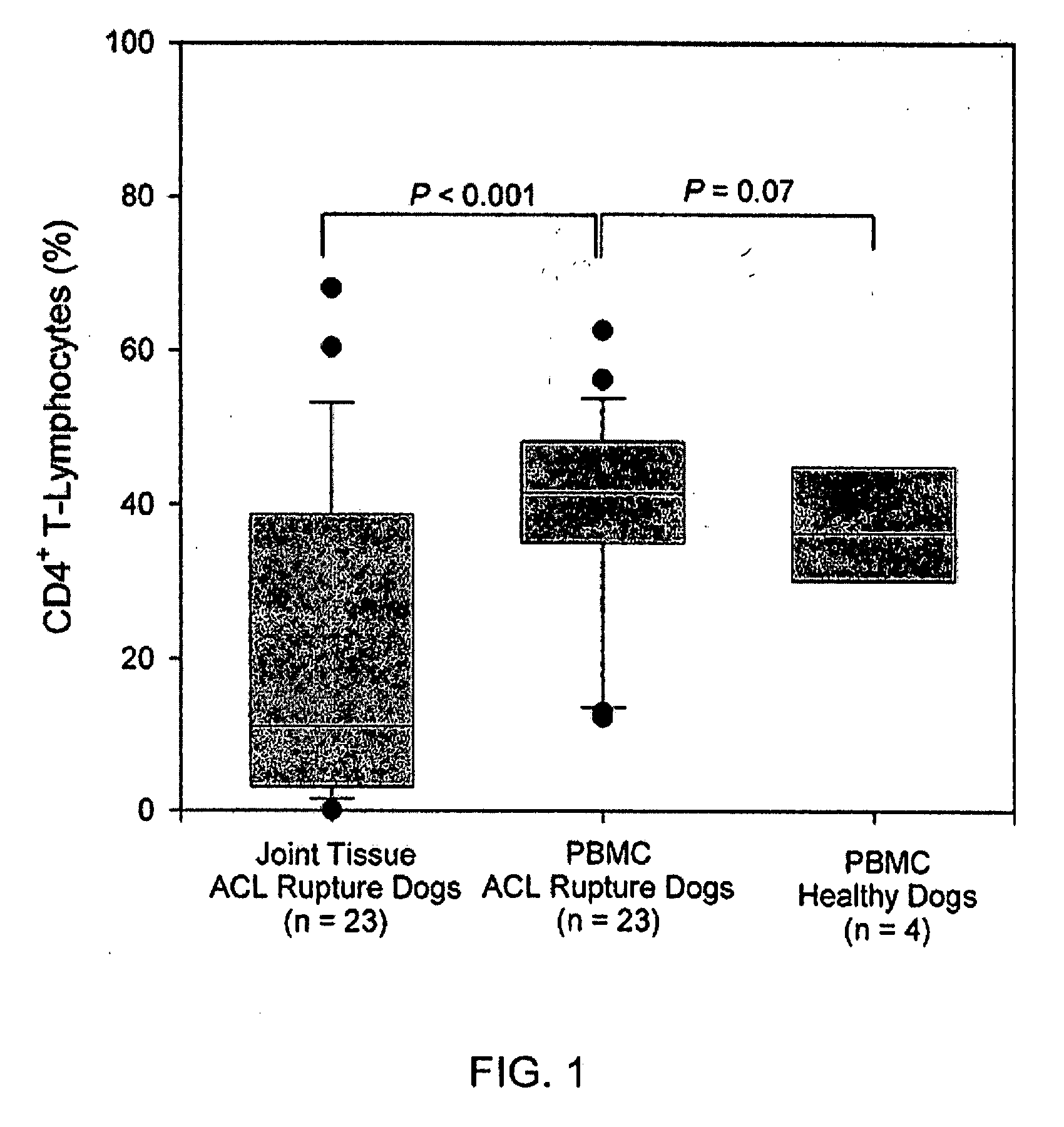
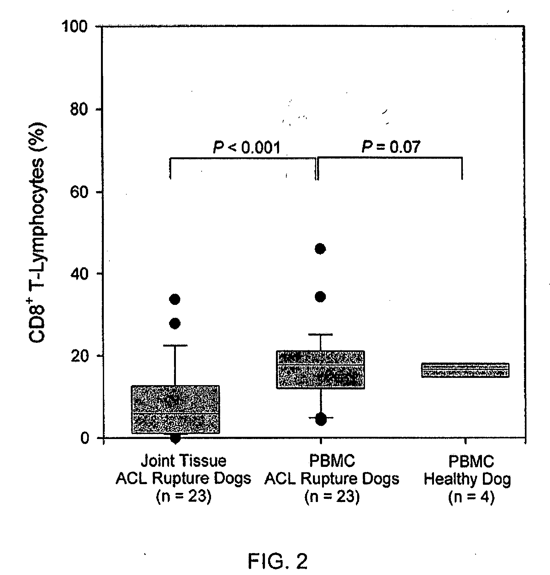
[0120] Blood and Joint Tissue Samples. Peripheral blood and specimens of synovium and ruptured ACL were collected from 23 dogs with inflammatory stifle arthritis / degenerative ACL rupture during surgical treatment. Peripheral blood samples were also collected from 4 healthy dogs with normal stifles. Procedures were conducted with the approval of the Animal Care Committee of the University of Wisconsin-Madison.

[0121] Preparation of PBMC for Flow Cytometry. PBMC were isolated using commercial cell separation tubes being BD Vacutainer™ CPT™, Becton Dickinson, Franklin Lakes, N.J.

[0122] After washing in phosphate buffered saline (PBS), erythrocytes were lysed. PBMC was then washed in RPMI with 10% fetal calf serum (RPMI / FCS). After determining the concentration of cells, cell counts were adjusted to ≦1×10⁷ / ml using FACS buffer. PBMC were then blocked and stained with fluorochrome-labeled anti-canine monoclonal antibodies directed against CD3, CD4, CD8, and CD44 epitopes, Serotec, Ralei...

example 2

[0142] Both synovial fluid and synovial membrane specimens were collected at surgery from a further 51 dogs with inflammatory stifle arthritis / degenerative ACL rupture. Dogs with PCR-positive joints were treated with doxycycline at 5 mg / kg bid orally for 10 weeks. Follow-up synovial fluid specimens were then collected aseptically by percutaneous needle aspiration.

[0143] Specimen collection and PCR for Bacterial DNA. Experiment #1. Synovial membrane specimens were collected aseptically during surgery. For the initial experiment, after extraction of DNA, a non-nested broad-range panbacterial PCR method was used for detection of DNA from the bacterial 16S rRNA gene. The PCR system used consensus primers for a highly conserved region of the gene. (Gerard H C et al., 2001).

[0144] All PCR reactions were performed in a laminar flow hood and extensive precautions were taken to prevent contamination of the PCR system. PCR products were examined under UV light after electrophoresis on a 1.5...

example 3

[0161] Blood and Joint Tissue Samples. Peripheral blood, synovium, and the ruptured ends of the CCL, when available, were collected from 23 dogs with stifle oligoarthritis and associated degenerative CCL rupture during surgical treatment. Age, weight, and gender were recorded for each dog. Peripheral blood and synovial tissues were also collected from 6 normal beagles with healthy stifle joints as a baseline for comparison with the dogs with oligoarthritis. The Beagles were all female; body weight and age were 10.5±1.7 kg and 3.3±3.0 years. Peripheral blood samples were also collected from five young healthy dogs of other breeds (Airedale Terrier, Pitbull Terrier, Toy Poodle, Gordon Setter and Boxer). The age and weight of these dogs were 21.5±9.8 kg and 1.5±1.4 years.

[0162] Preparation of Peripheral Blood Mononuclear Cells (PBMC) for Flow Cytometry. PBMC were isolated using commercial cell separation tubes (BD Vacutainer™ CPT™, Becton Dickinson, Franklin Lakes, N.J.). After washin...


PUM

PropertyMeasurementUnit
Electrical conductanceaaaaaaaaaa
Concentrationaaaaaaaaaa
Degradation propertiesaaaaaaaaaa

Abstract

Method of diagnosing persistent, chronic synovitis and progressive joint degradation in the joint of a mammal. The method including providing a test sample comprising synovial fluid, cell or tissue from the joint of a mammal; detecting the presence of bacterial DNA, measuring the concentration of one or more biomarkers being cathepsin K, MMP-2 and -9, cathepsin S, tartrate-resistant acid phosphatase, invariant chain, CD4+ T-lymphocytes, CD8+ T-lymphocytes, CD44+ mononuclear cells, Toll-like receptor-2, Toll-like receptor-9, or a combination thereof; and, comparing the concentration of the biomarker from the test sample to a corresponding biomarker concentration in a control sample from healthy dogs, or an internal PBMC control sample, wherein a statistically significant elevated concentration of the biomarker in the test sample indicates that the mammal's joint is diseased.

Description

CROSS-REFERENCE TO RELATED APPLICATIONS [0001] This application claims priority to U.S. Provisional Patent Application Ser. No. 60 / 838,469 filed on Aug. 17, 2006. [0002] U.S. Patent Application Publication No. US 2005 / 0074800 A1 was published on Apr. 7, 2005, which is incorporated herein by reference.FIELD OF THE INVENTION [0003] The field of the invention relates to kits and methods for detecting synovial disease in the knee of a human or the stifle dog. The anatomically correct term is “knee” for humans and “stifle” for dogs. BACKGROUND OF THE INVENTION [0004] It has been reported that in the US more than $1 billion is spent annually treating ruptured ligaments in dogs. It has been generally assumed that such ruptures are due to trauma or natural tissue degeneration. However, increasing evidence suggests that there may be an immunological component to such ligament disease. That link is based on findings showing large amounts of macrophages, dendritic cells, and T lymphocytes in t...

Claims


Application Information

Patent Timeline
IPC IPC(8): A61K31/65A61P19/02C12Q1/02C12Q1/42C12Q1/68G01N33/48G01N33/68
CPCC12Q1/6883C12Q2600/158Y10T436/143333G01N2800/10G01N2800/102G01N33/6893A61P19/02Y02A50/30
Inventor MUIR, PETER
Owner WISCONSIN ALUMNI RES FOUND
Why Eureka
Try Eureka