Eureka AIR delivers breakthrough ideas for toughest innovation challenges, trusted by R&D personnel around the world.

Micro vein enhancer

a vein enhancer and micro-vein technology, applied in the field of micro-vein enhancers, can solve the problems of relativly difficult design and build a system that closely aligns the captured image and the projected image, and achieves the effect of reducing and/or reducing the amount of botched attempts to pierce a vein and being convenient to opera

Active Publication Date: 2007-07-12
ACCUVEIN
View PDF100 Cites 44 Cited by
  • Summary
  • Abstract
  • Description
  • Claims
  • Application Information

AI Technical Summary

Benefits of technology

[0008]The present invention is a Miniature Vein Enhancer that includes a Miniature Projection Head and a mounting means for the Miniature Projection head. The Miniature Projection Head of the present invention implements a polarized laser light. This diminishes the effects of specular reflection off the surface of the skin. The Veinviewer Imaging System, produced by Luminetx, uses a polarized filter to polarize the LED light. This polarized LED light is then rotated 90° in front of the camera, thus causing increased power loss. In addition, the IR and visible lasers in the present invention are modulated to allow a regular photodiode to detect the different signals from each wavelength separately. Furthermore, the IR laser power of the present invention is dynamically altered during each scan line, thus increasing the working range of the photodiode, and allowing for constant DC gain.
[0009]The miniature vein enhancer of the present invention may be used by a practitioner to locate a vein, particularly useful when trying to locate a vein in the very old or very young. More then fifty percent of attempts to find a vein in old people, who have a generally high percentage of loose, fatty tissue, and children, who have a generally high percentage of small veins and “puppy fat” are unsuccessful. The present invention is aimed at reducing and / or preventing the discomfort and delay associated with botched attempts to pierce veins for injections and blood tests. In addition, the present invention can cut the time it takes to set up potentially life-saving intravenous drip.OBJECTS OF THE INVENTION
[0011]It is another object of the present invention to make a Miniature Vein Enhancer that will allow a practitioner pinpoint a vein for intravenous drip, blood tests, and the like.
[0012]It is still another object of the present invention to make a Miniature Vein Enhancer that will reduce and / or diminish the amount of botched attempts to pierce a vein.
[0013]It is still a further object of the present invention to make a Miniature Vein Enhancer that is easy to operate.

Problems solved by technology

Given that the CCD imager and the image projector are both two dimensional, and do not occupy the same point in space, it is relatively difficult to design and build a system that closely aligns the captured image and the projected image.

Method used


Image

  • Micro vein enhancer
  • Micro vein enhancer
  • Micro vein enhancer

Examples


Embodiment Construction

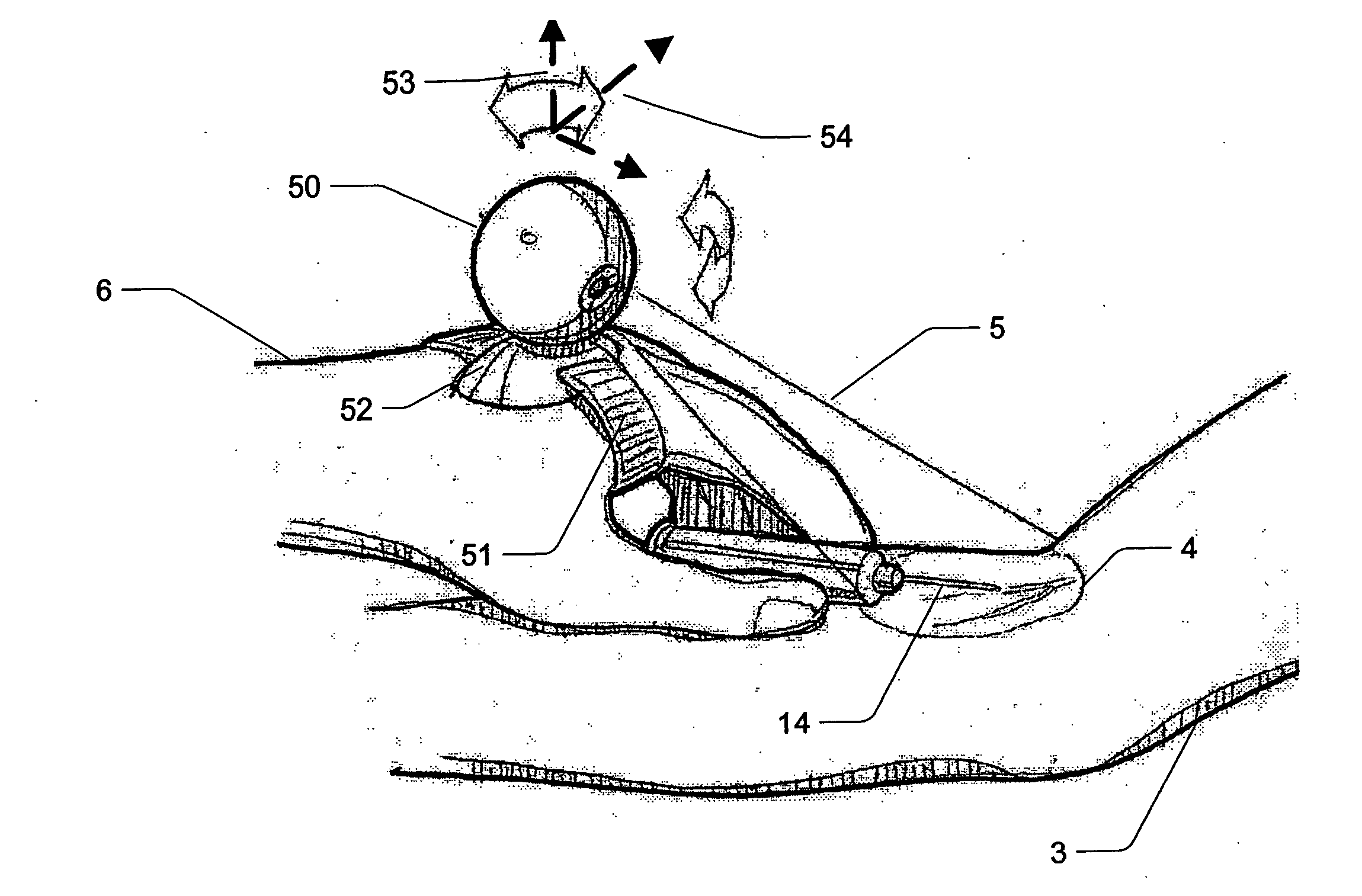
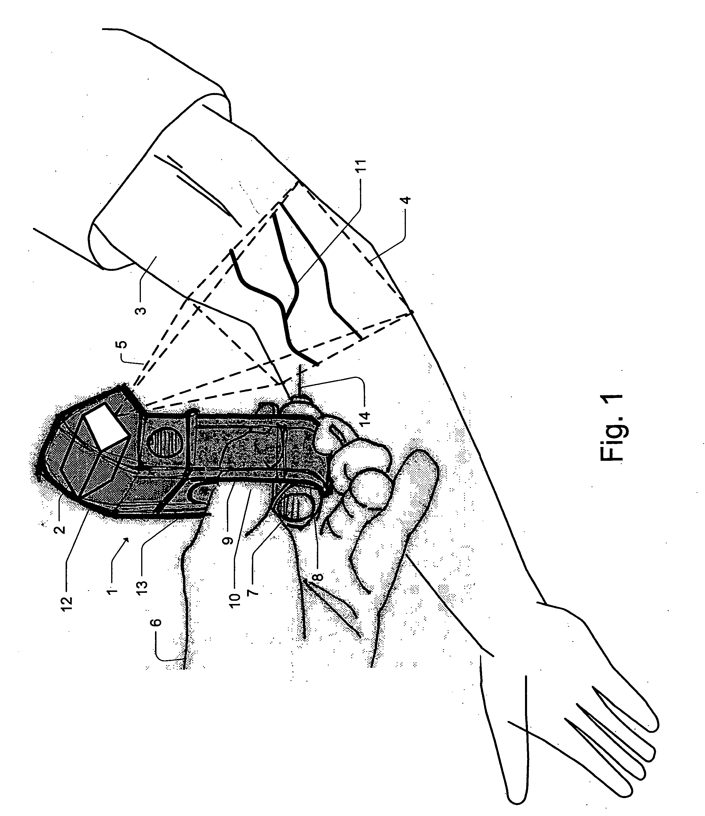
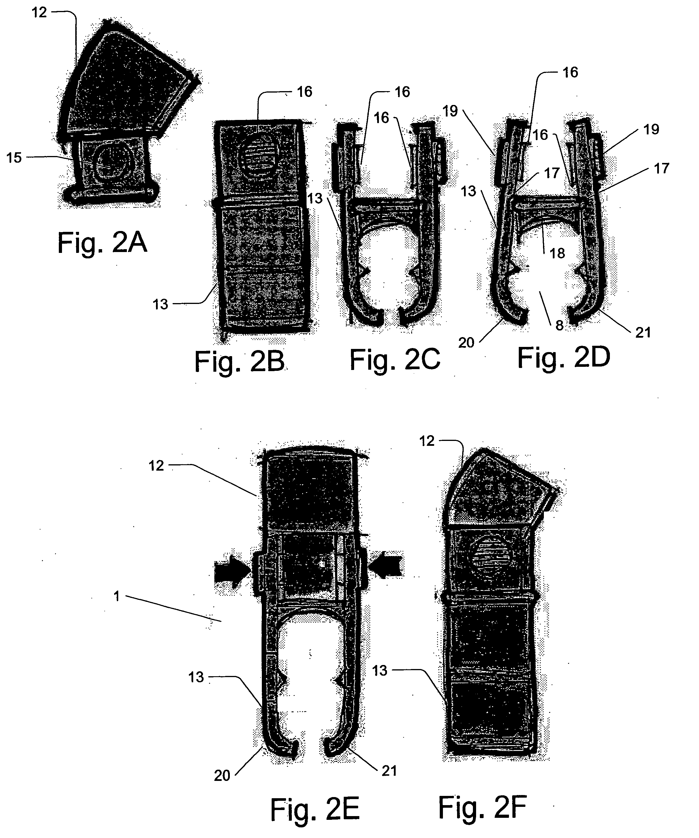
[0100]FIG. 1 shows a miniature vein enhancer (MVE) 1 for enhancing a target area 4 of a patient's arm 3. The MVE 1 has miniature projection head (MPH) 2 for both imaging the target area 4 and for projecting an enhanced image 11 along optical path 5 onto the target area 4. The MPH will be described in detail later with reference to FIG. 18-FIG. 21. The MPH 2 is housed in a cavity section preferably a top cavity section 12 of the MVE 1. The body 13 of the MVE 1 is positioned below the top cavity section 12. The body 13 has a vial opening 8 for receiving and temporarily holding in place a vial holder 7 having a needle 14. The body 13 also has a thumb opening 9 through which the medical practitioner 6 can place their thumb 10 while utilizing the MVE 1. The vial opening 8 is preferably provided with at least a curved base section 8A for receiving the curved exterior surface of the vial holder 7 and retaining it in position. The thumb opening 9 may be a separate orifice or it may be part ...


PUM


Abstract

The present invention is a Miniature Vein Enhancer that includes a Miniature Projection Head. The Miniature Projection Head may be operated in one of three modes, AFM, DBM, and RTM. The Miniature Projection Head of the present invention projects an image of the veins of a patient, which aids the practitioner in pinpointing a vein for an intravenous drip, blood test, and the like. The Miniature projection head may have a cavity for a power source or it may have a power source located in a body portion of the Miniature Vein Enhancer. The Miniature Vein Enhancer may be attached to one of several improved needle protectors, or the Miniature Vein Enhancer may be attached to a body similar to a flashlight for hand held use. The Miniature Vein Enhancer of the present invention may also be attached to a magnifying glass, a flat panel display, and the like.

Description

[0001]This application claims priority from previously filed provisional application 60 / 757,704, entitled Micro Vein Enhancer, filed on Jan. 1, 2006, all disclosures of which are hereby incorporated by reference.FIELD OF INVENTION[0002]A miniature laser based vein contrast enhancer that can fit into portable hand held products that a practitioner can carry in their pocket.BACKGROUND OF THE INVENTION[0003]It is known in the art to use an apparatus to enhance the visual appearance of the veins in a patient to facilitate insertion of needles into the veins. An example of such a system is described in U.S. Pat. Nos. 5,969,754 and 6,556,858 incorporated herein by reference as well as a publication entitled “The Clinical Evaluation of Vein Contrast Enhancement”. Luminetx is currently marketing such a device under the name “Veinviewer Imaging System” and information related thereto is available on its website, which is incorporated herein by reference.[0004]The Luminetx Vein Contrast Enhan...

Claims


Application Information

Patent Timeline
Patent Type & Authority Applications(United States)
IPC IPC(8): A61B6/00
CPCA61B5/0059A61B5/489A61B2019/5293A61M5/427A61B5/0037A61B5/743A61B5/0082A61B5/7475A61M5/46A61B5/0068A61B5/0079A61B5/0075A61B2090/366A61B90/13A61M5/42A61B5/00G06T7/50G02B27/1006A61B5/0062A61B5/0071A61B5/6824A61B5/6831A61B2560/0431A61B5/6887A61B17/3403A61B2560/0214A61B2560/0425A61B2562/028
Inventor GOLDMAN, RONHUNT, DAVIDMOCK, MARKMARSHAL, GRAHAMCONLON, STEPHEN P.ROTH, BOB
Owner ACCUVEIN
Why Eureka
Eureka Blog
Learn More