
Methods of rapid chromatin immunoprecipitation

a chromatin immunoprecipitation and chromatin technology, applied in the field of rapid chromatin immunoprecipitation, can solve the problems of time-consuming and time-consuming current chip method, cannot be used for analyzing dna-protein interactions in living cells, and achieve rapid and efficient chip, rapid and convenient binding, and rapid reverse crosslinked protein-dna complex

Inactive Publication Date: 2007-06-21
LI WEIWEI +1
View PDF3 Cites 16 Cited by
  • Summary
  • Abstract
  • Description
  • Claims
  • Application Information

AI Technical Summary

Benefits of technology

The present invention provides a method and kit for quickly identifying regions of the genome that specific proteins bind to, or identifying specific proteins that bind to a specific region of the genome in vivo. This is achieved by crosslinking protein to DNA in living cells or fresh tissues with formaldehyde, and then capturing the protein-DNA complex with affinity antibodies. The method is efficient and effective, and can be performed at room temperature. The invention overcomes weaknesses in previous technologies and provides a faster and more reliable method for identifying protein-DNA interactions.

Problems solved by technology

However these technologies can not be used for analyzing DNA-protein interactions in a living cell, which is more a complex environment than that in vitro.
However the currently used CHIP method has several drawbacks.
The most critical weakness of the current CHIP method is time consuming.
In addition, the current CHIP method uses suspended beads to capture antibody / complex and centrifugation-dependent wash, which makes the procedure to be labor-intensive and to have low throughput.

Method used


Image

  • Methods of rapid chromatin immunoprecipitation
  • Methods of rapid chromatin immunoprecipitation
  • Methods of rapid chromatin immunoprecipitation

Examples


example 1

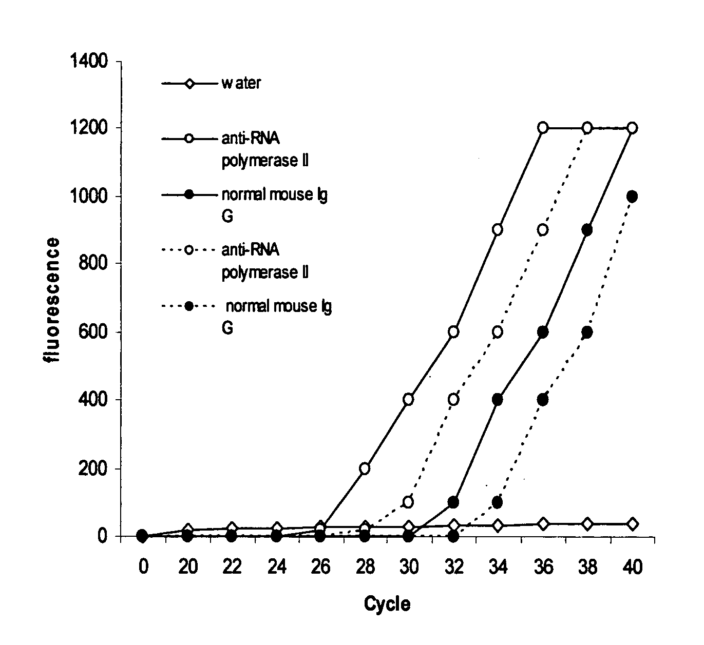
[0030]The experiment was carried out to detect whether a specific protein binds to the specific sequences of a gene in living cells.

[0031]Antibody binding to the assay plate: Wash plate wells twice with wash buffer (100 mM sodium phosphate, 200 mM NaCl, pH 7.4 and 1% Triton X 100). Dilute various antibodies with wash buffer to 10 μg per ml and add 100 μl of diluted antibody to the each well. Normal mouse Ig G was used as the negative control and anti-RNA polymerase II as the positive control. Cover the plate and incubate at room temperature for 60-90 min. The plate was then washed 3-4 times with wash buffer.

[0032]Cell fixation and lysis: Hela cells were grown to 70%-80% confluence on a 100 mm plate, then trypsinized and collected into a 15 ml conical tube. After washing with PBS, cells were suspended in 9 ml of fresh culture medium containing 1% formaldehyde (final concentration) and incubated at room temperature (20-25° C.) for 10 min on a rocking platform (50-100 rpm). 1 ml of gly...

example 2

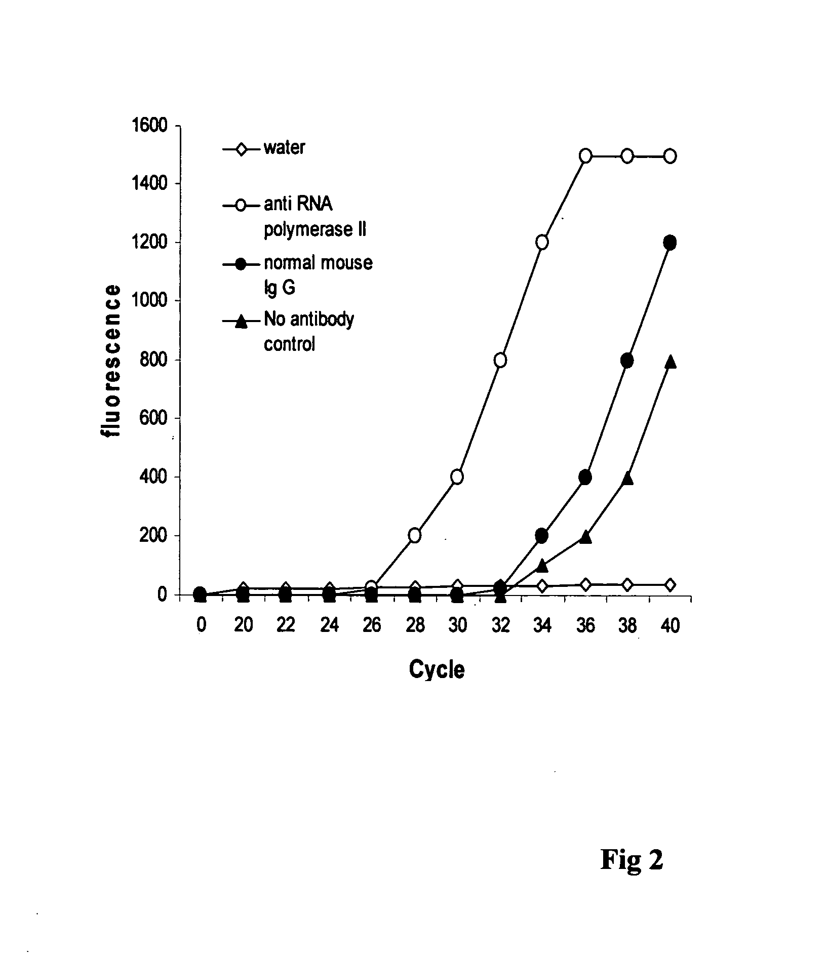
[0037]The experiment was carried out to compare the effect of the CHIP method of this invention to that of conventional CHIP.

[0038]Antibody binding to the assay plate: Wash plate wells twice with wash buffer (100 mM sodium phosphate, 200 mM NaCl, pH 7.4 and 1% Triton X 100). Dilute various antibodies with wash buffer to 10 μg per ml and add 100 μl of diluted antibody to the each well. Normal mouse Ig G was used as the negative control and anti-RNA polymerase II as the positive control. Cover the plate and incubate at room temperature for 60-90 min. The plate was then washed 3-4 times with wash buffer.

[0039]Hela cells were grown to 70%-80% confluence on a 100 mm plate, then trypsinized and collected into a 15 ml conical tube. After washing with PBS, cells were suspended in 9 ml of fresh culture medium containing 1% formaldehyde (final concentration) and incubated at room temperature (20-25° C.) for 10 min on a rocking platform (50-100 rpm). 1 ml of glycine (1.25 M) was added and incu...


PUM

PropertyMeasurementUnit
temperatureaaaaaaaaaa
concentrationaaaaaaaaaa

Abstract

This invention is related to a method for rapidly identifying regions of the genome to which specific proteins bind, or identifying specific proteins bound to a region of the genome in vivo.

Description

CROSS-REFERENCE TO RELATED APPLICATIONS[0001]Not applicableSTATEMENT REGARDING FEDERALLY SPONSORED RESEARCH OR DEVELOPMENT[0002]Not applicableREFERENCE TO A MICROFICHE APPENDIX[0003]Not applicableBACKGROUND OF THE INVENTION[0004]1. Field of the Invention[0005]This invention is related to a method for rapidly identifying regions of the genome to which specific proteins bind, or identifying specific proteins bound to a region of the genome in vivo.[0006]2. Description of the Related Art[0007]Protein-DNA interactions are widely involved in a variety of molecular processes of living cells such as single transduction, gene transcription, chromosome segregation, DNA replication and recombination, and epigenetic silencing of genes (Ptashne M and Cann A. Genes and signals, Cold spring Harbor laboratory Press, 2001, Das, P. M., Ramachandran, K. venWart, J., Signal, R. Biotechniques, 37: 961-969, 2004.). Identifying the genetic targets of DNA binding proteins and knowing the mechanisms of pro...

Claims


Application Information

Patent Timeline
Patent Type & Authority Applications(United States)
IPC IPC(8): C12Q1/68C12N1/08
CPCC12Q1/6806C12Q2527/125C12Q2527/101
Inventor LI, WEIWEILI, JESSICA M.
Owner LI WEIWEI
Learn More