
Method and x-ray apparatus for exposure of a patient who can be placed at a variable distance relative to an x-ray source

a patient and variable distance technology, applied in the field of method and an x-ray apparatus for radiation exposure of a patient, can solve the problems of unacceptably high dose rate of ionizing radiation, unacceptably high entrance dose rate of patient, and patient lying on the table being irradiated with unacceptably high dose ra

Inactive Publication Date: 2006-11-30
SIEMENS HEALTHCARE GMBH
View PDF6 Cites 18 Cited by
  • Summary
  • Abstract
  • Description
  • Claims
  • Application Information

AI Technical Summary

Benefits of technology

[0015] An object of the present invention is to provide a method and a corresponding x-ray apparatus for fluoroscopy of a patient who can be placed at a variable distance relative to an x-ray source wherein the aforementioned problems are avoided or made less severe.
[0018] This method therefore ensures that the patient is never exposed with a dose rate that is too high. The x-ray imaging can also be continued at distances between the patient and the x-ray source that lie below the safe distance for the maximum output capacity of the x-ray source, without exceeding the limit dose rate, i.e., the maximum allowed dose rate that strikes the patient. Although the image quality is then possibly limited, imaging still occurs without having to move the patient again away from the x-ray source and position that is ergonomic for the doctor can be achieved.
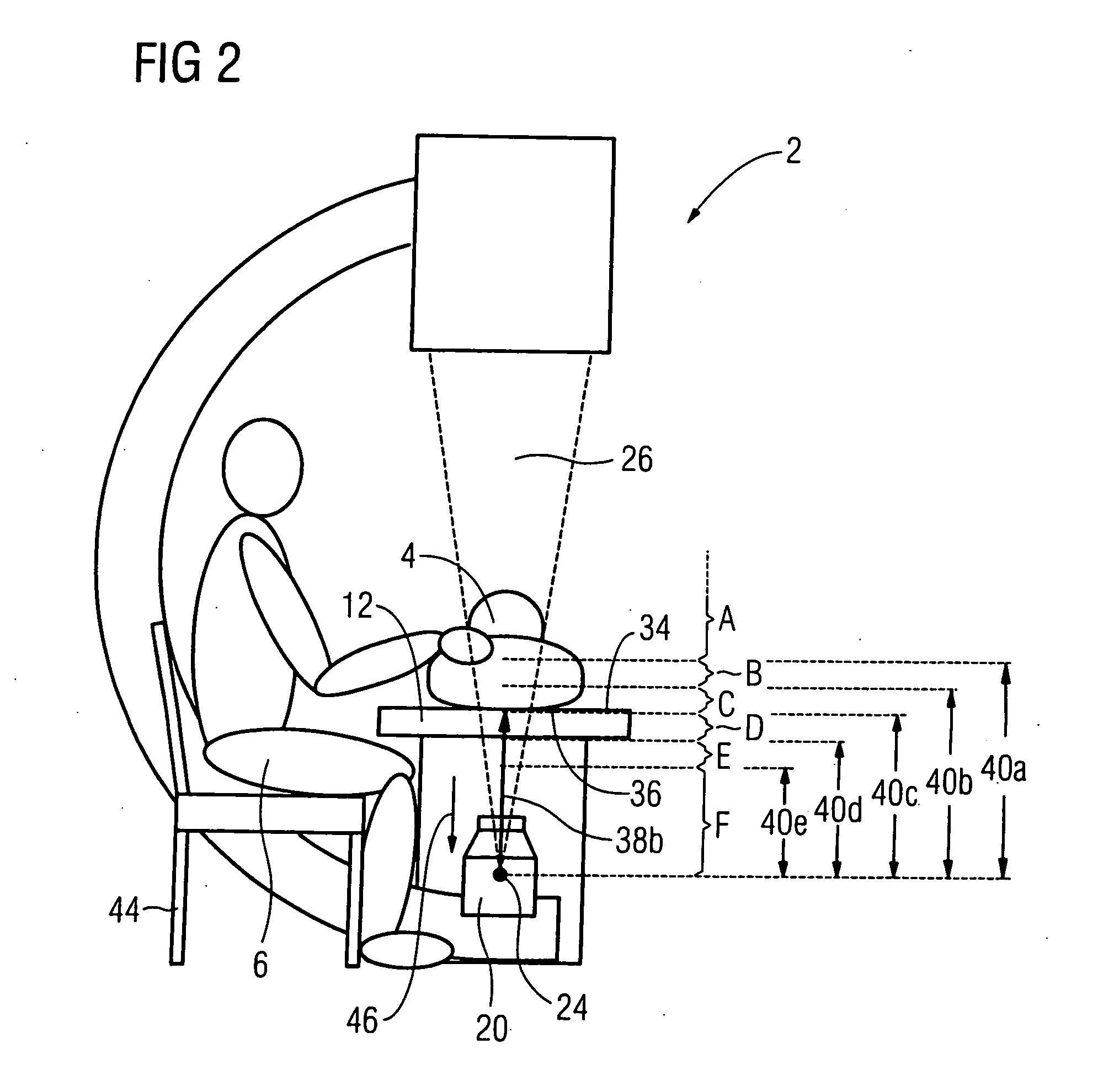
[0021] Alternatively, the dose rate can be varied in steps given specific interval limits of the distance between the patient and the x-ray source. For example, an interval limit exists that is associated with the maximal emittable power of the x-ray source. For intervals (spacings) above this interval limit, the patient is in no way irradiated with a dose rate that is too high, even when the x-ray source radiates with its system-dependent maximum dose rate. Moreover, further interval limits can be predetermined that are respectively associated other reduced output capacities of the x-ray source. The output power radiated by the source is then constant between two interval limits such that, for example, the remaining components of the imaging system can likewise be adjusted in steps to the respective source characteristic. Complicated, continuous updating tracking of the component properties is thus avoided.
[0022] The variation of the dose rate upon the distance (the current distance) approaching an interval limit can be terminated beforehand. A person implementing the method thus has discretion to leave a patient (for example) slightly above or slightly below a specific interval limit in order to avoid a reduction of the dose rate emitted by the x-ray source, so as to not impair the image quality of x-ray exposures. A possible decisive advantage in image quality can thus be achieved through a slight loss in ergonomic comfort.
[0023] The dose rate can be automatically varied. The implementing person thus does not have to attend to the adherence to safety intervals or the like since, for example, the dose rate is automatically adapted given free selection of the patient distance from the x-ray radiator or a suitable patient distance is set given prior selection of a specific image quality. For example, in a critical phase of an operation the doctor can thus automatically attain an x-ray image with improved image quality simply by raising the treatment table.
[0026] The method can be executed particularly simply when the filter is brought between the x-ray source and the patient in a motorized manner. The entire method thus can be automated with no personnel for manual pivoting being necessary.

Problems solved by technology

Despite this large usage in medicine, such ionizing radiation is not free of health risks for the patient.
For the most part, the patient table can be moved toward the x-ray radiator so far that, upon operation of the x-ray radiator, the patient lying on the table would be irradiated with an unacceptably high dose rate.
The minimum distance may then no longer be adhered to, which (given an activated x-ray source) would lead to an unacceptably high entrance dose rate at the patient.
The radiation, however, is hardened by the primary aluminum filter, causing the image noise in the x-ray image to increase in a disruptive manner, and in fact at distances between the patient and the x-ray source that would be non-critical given unattenuated x-ray radiation (without an aluminum filter).
Furthermore, it is known to mechanically limit the movement path of the patient table in a fixed manner such that thus cannot be moved closer to the x-ray source than is prescribed by the safety interval.
This limits the ergonomics of the arrangement since the table is precluded from being lowered when the urologist would like to execute a procedure without x-ray exposure.
In such a table position, however, x-ray fluoroscopy of the patient is no longer possible at all.
This likewise leads to a permanent decrease of the maximum safety distance.
As in the case of a permanently-installed filter, however, the image quality of the x-ray system is generally degraded since the x-ray source is never operated at its optimal operating point.
The decrease of the generator power leads to a decrease of the number of generated x-ray quanta and an increase of the quantum noise.
The x-ray image is coarser, appears noisy and is no longer regarded as being acceptable.
All known solutions are thus compromises between the x-ray contrast or the x-ray image quality and the ergonomics of the overall arrangement.

Method used


Image

  • Method and x-ray apparatus for exposure of a patient who can be placed at a variable distance relative to an x-ray source
  • Method and x-ray apparatus for exposure of a patient who can be placed at a variable distance relative to an x-ray source
  • Method and x-ray apparatus for exposure of a patient who can be placed at a variable distance relative to an x-ray source

Examples


Embodiment Construction

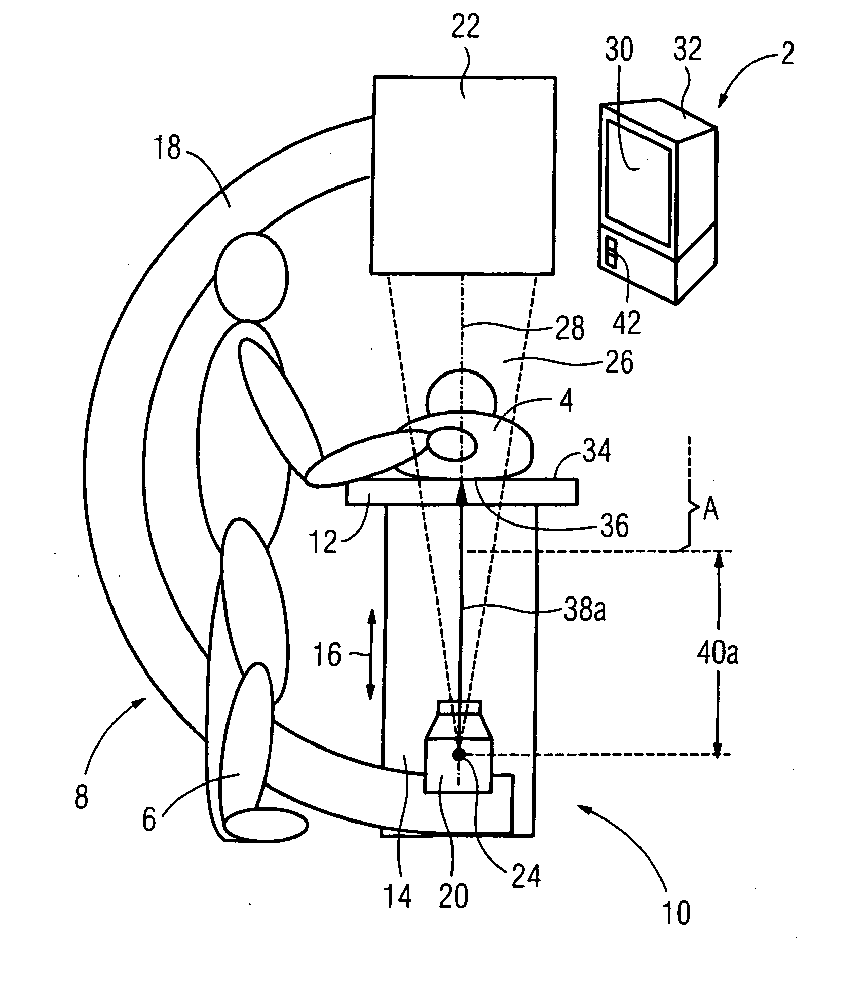
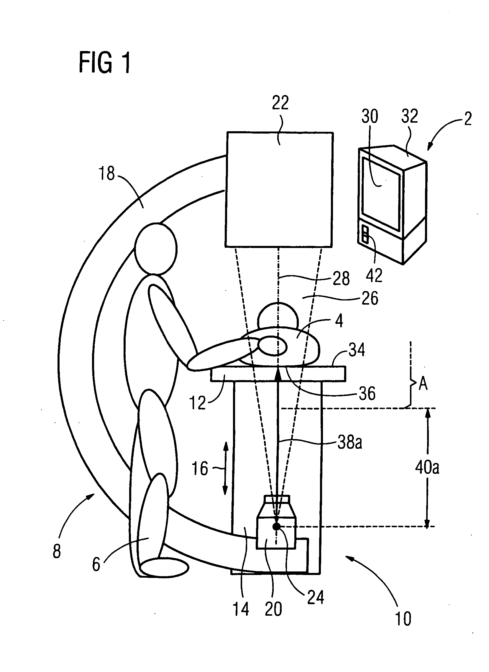
[0041]FIG. 1 shows a urological workstation 2 with a patient 4 and a doctor 6. The workstation 2 has an x-ray C-arm unit 8 and a patient table 10 with a table plate 12 on which the patient 4 lies. The table plate 12 is supported on a base 14 such that it can be adjusted in terms of height in the direction of the double arrow 16. The height adjustment ensues by actuation of a control (operating) lever 42 in a manner that is not necessary to explain in detail.

[0042] The x-ray C-arm unit 8 has a C-shaped supporting arm 18 at the ends of which an x-ray source 20 and an x-ray image intensifier 22 are mounted, respectively. In the x-ray source 20 (more precisely, the x-ray tube contained therein and not shown), x-ray radiation 26 (represented in FIG. 1 by a beam cone) is emitted from the focus 24 along a central ray 28 toward the x-ray image intensifier 22. The x-ray radiation 26 penetrates the patient 4 in order to generate an x-ray image 30 of the inside of the body in a known manner, ...


PUM


Abstract

In a method and x-ray apparatus for exposure of a patient who can be placed at a variable distance relative to an x-ray source, the dose rate emitted by the x-ray source is reduced given reduction of the distance and / or increased given increase of the distance.

Description

BACKGROUND OF THE INVENTION [0001] 1. Field of the Invention [0002] The present invention concerns a method and an x-ray apparatus for radiation exposure of a patient who can be placed at a variable distance relative to an x-ray source. [0003] 2. Description of the Prior Art [0004] X-ray methods and x-ray apparatuses have been used for many decades for medical imaging, in which x-rays are directed through the body of a patient in the form of a living person or animal. Despite this large usage in medicine, such ionizing radiation is not free of health risks for the patient. The use of x-ray radiation on living patients is therefore heavily legally regulated. In particular, maximal allowable limit values exist for the dose rate of x-ray radiation on the body surface of a patient. 21 C.F.R. (Code of Federal Regulations) of the Department of Health and Human Services is an example. 21 C.F.R. §1020.32e specifies what is known as the maximum allowed input exposure rate, thus the entrance ...

Claims


Application Information

Patent Timeline
IPC IPC(8): A61N5/10
CPCA61B6/0457A61B6/06A61B6/589A61B6/542A61B6/4441A61B6/0487
Inventor FEHRE, JENSKALTSCHMIDT, RAINERLUDWIG, MARKUSWILL, ULRICH
Owner SIEMENS HEALTHCARE GMBH
Learn More