
Method for detecting infectious agents using computer controlled automated image analysis

an image analysis and computer controlled technology, applied in the field of computer controlled methods and apparatus for detecting or detecting and quantifying infectious agents, can solve the problems of increasing the risk of heart disease, triggering an inflammatory response in the vessel wall,

Inactive Publication Date: 2006-10-26
IKONISYS INC
View PDF28 Cites 24 Cited by
  • Summary
  • Abstract
  • Description
  • Claims
  • Application Information

AI Technical Summary

Benefits of technology

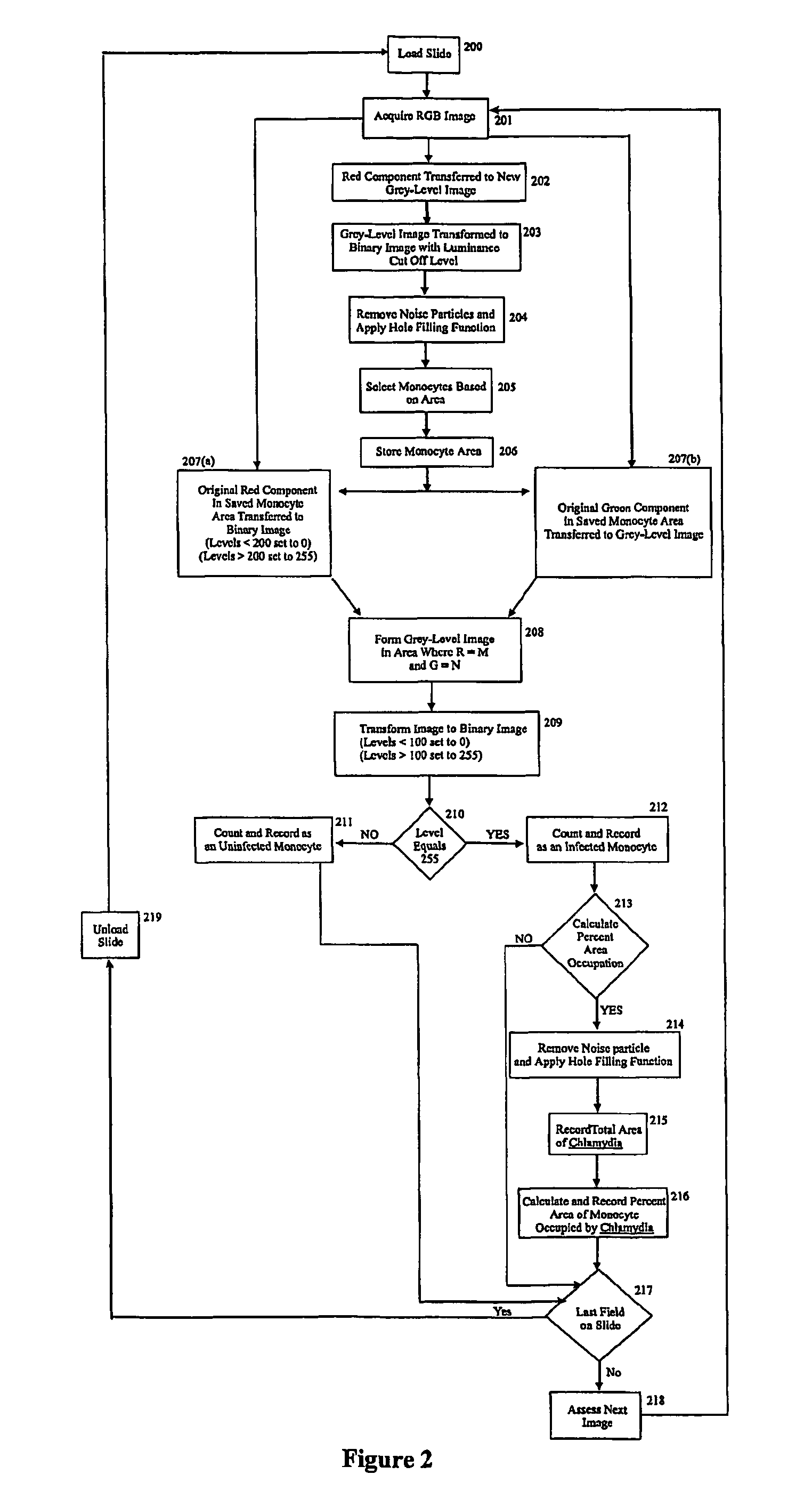
[0016] In an embodiment, the image data is transformed from one color space, e.g., an RGB (Red Green Blue) image into another color space, e.g., HLS (Hue Luminescence Saturation) image. Filters and / or masks are utilized to distinguish those cells that meet pre-selected criteria, i.e. contain a detectable signal, and eliminate those that do not, and thus identify infected animal cells.
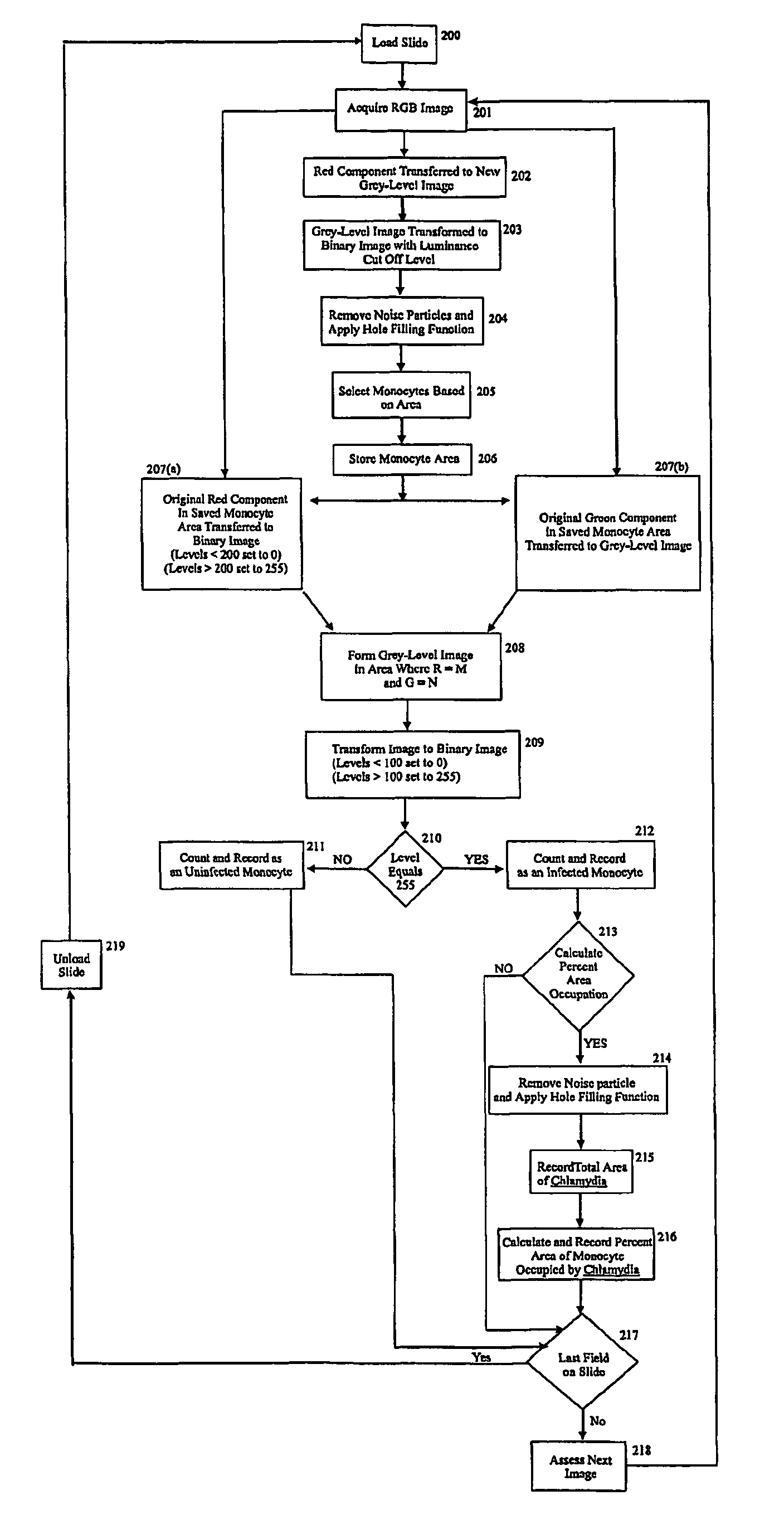
[0023] The Red component of the RGB image is transferred to a new monochrome grey-level image and clipped for pixel values of less than 50 to cut down signal noise.
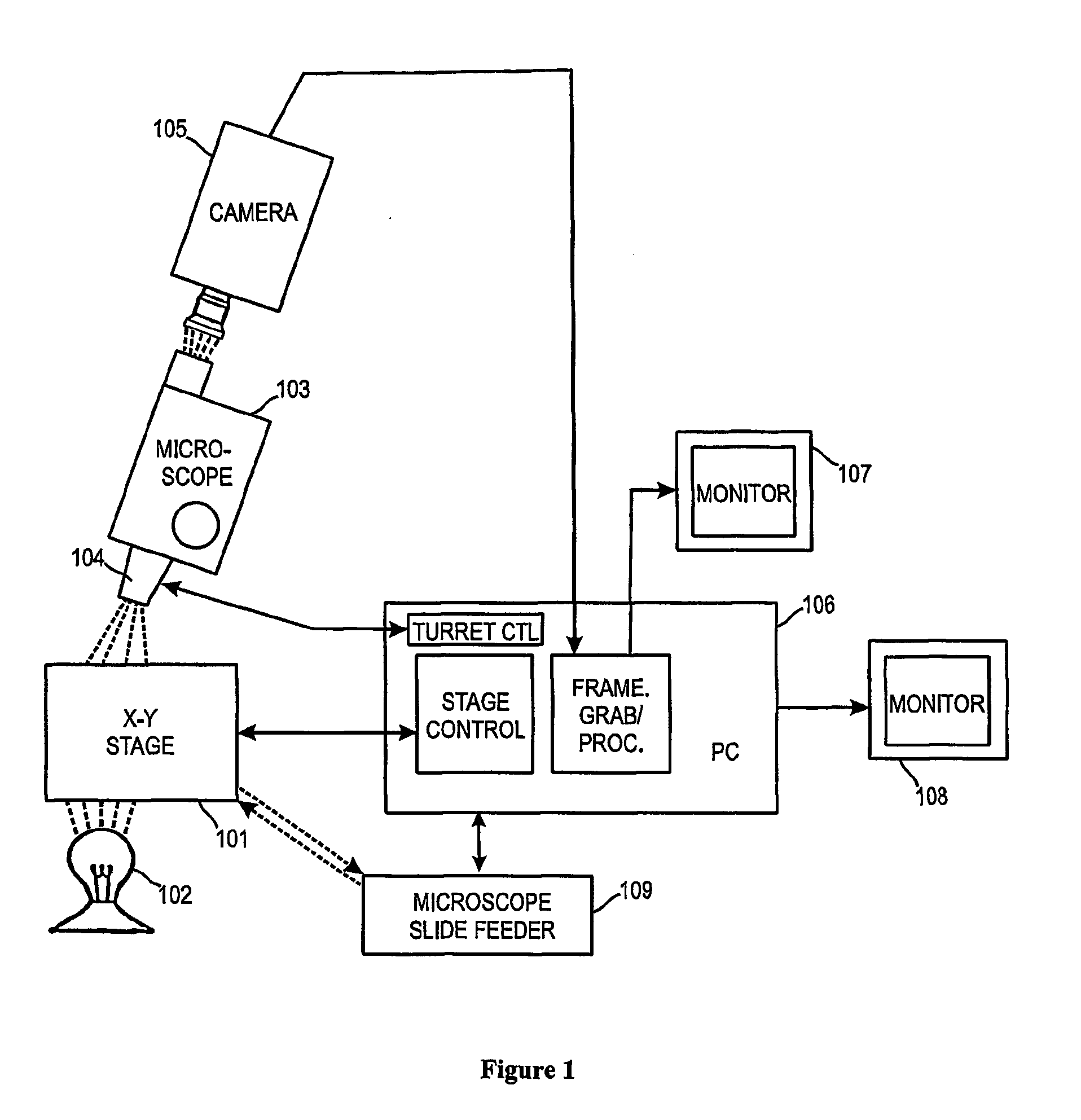
[0056] The present invention also provides a system for screening infected animal cells or infected body fluids or tissues. The basic elements of the system include an X-Y stage, a mercury light source, a fluorescence microscope, digital camera system such as a color CCD camera, or a complementary metal-oxide semiconductor (CMOS) image system, a personal computer (PC) system, and one or two monitors and most importantly a computer software product including a computer-readable storage medium having fixed therein a sequence of instructions which, when executed by a computer, direct the performance of steps for conducting the methods of the invention as described herein. In a preferred embodiment, the stage has an automated microscope feeder configured with it so that slides can be automatically moved into and out of position for image capture and / or analysis.

Problems solved by technology

Many systemic and local factors may cause dysfunctional endothelium and lead to or trigger an inflammatory response in the vessel wall.
Studies have shown Herpes viruses, such as Herpes simplex virus and Cytomegalovirus (“CMV”), can increase the risk of developing heart disease.

Method used


Image

  • Method for detecting infectious agents using computer controlled automated image analysis
  • Method for detecting infectious agents using computer controlled automated image analysis
  • Method for detecting infectious agents using computer controlled automated image analysis

Examples


Embodiment Construction

[0068] The invention will be better understood upon reading the following detailed description of the invention and of various exemplary embodiments of the invention, in connection with the accompanying drawings. It will be clear to those skilled in the art that the invention can be applied to and, in fact, encompasses methods and systems to provide information useful in making a disease diagnosis or a prognosis of disease or disease susceptibility or outcome based on detection and preferably, quantification of any characteristic resulting from infection of a host, e.g., an animal cell with an infectious agent. The animal cell can be obtained from any fluid or tissue sample from which it is possible to view a monolayer of cells infected with said infectious agent. It will also be clear to those skilled in the art that the invention can be applied to, and in fact, encompasses methods and systems to provide information useful in making a disease diagnosis or prognosis of disease or di...


PUM

PropertyMeasurementUnit
vascular disorderaaaaaaaaaa
optical fieldaaaaaaaaaa
sizeaaaaaaaaaa

Abstract

Computer controlled methods, systems and apparatus for detecting an infectious agent in a host cell, an animal cell, or plant cell, a body fluid or tissue sample to provide information useful to diagnose a disease or prognosticate disease susceptibility, extent or outcome are provided. In one illustrative embodiment, the infected animal cell is a Chlamydia infected mononuclear phagocyte in a blood sample. In another illustrative embodiment, the infected body fluid is a blood sample infected with “fee floating”Chlamydia. Detection and preferably, quantification of such an infected mononuclear phagocyte(s) or blood sample provides information useful in diagnosing or prognosticating susceptibility, extent or outcome of patients suspected of suffering from vascular, coronary or central nervous disease(s) or disorder(s).

Description

[0001] This application claims priority benefits of U.S. Patent Application No. 60 / 295,587 filed Jun. 4, 2001, the entire disclosure of which is incorporated herein by reference.1. FIELD OF THE INVENTION [0002] The present invention relates to computer controlled methods and apparatus for detecting or detecting and quantifying an infectious agent in a biological sample. The identifying and / or quantitative data obtained are useful in making a diagnosis or prognosis of diseases, including diseases classically deemed “non-infectious.” In one aspect, the invention relates to computer controlled methods and systems for identifying an infected host, animal or plant cell in a field of cells and thereby providing information useful in making a disease diagnosis or a prognosis of disease susceptibility based on identification of an infected animal cell. In another aspect, the invention relates to computer controlled methods and systems for quantification of infected host, e.g., animal or pla...

Claims


Application Information

Patent Timeline
Patent Type & Authority Applications(United States)
IPC IPC(8): G06M11/02G01N33/48G06K9/00A61BG06T7/00
CPCC12Q1/04G01N15/1475G06K9/00147G06T7/0012G06T2207/30024G06K9/00127G06V20/69G01N15/1433G06V20/698
Inventor TAFAS, TRIANTAFYLLOSKILPATRICK, MICHAELTSIPOURAS, PETROS
Owner IKONISYS INC
Learn More