
Novel field testing method and device for detection of equine protozoal myeloencephalitis

a protozoal and myeloencephalitis technology, applied in the field of field testing methods and devices for detection of equine protozoal myeloencephalitis, can solve the problems of insufficient understanding of the disease, insufficient use of diagnostic tests, and inability to achieve significant understanding, diagnosis and treatment of the diseas

Inactive Publication Date: 2006-09-07
KRISTIPATI RAMA SHARMA +3
View PDF2 Cites 0 Cited by
  • Summary
  • Abstract
  • Description
  • Claims
  • Application Information

AI Technical Summary

Benefits of technology

[0030] To achieve the foregoing and other objects and in accordance with the purpose of the present invention as embodied and broadly described herein, the present invention is directed to a quick, inexpensive immuno-diagnostic method for the detection of a specific pathogen in body fluids of humans and animals.
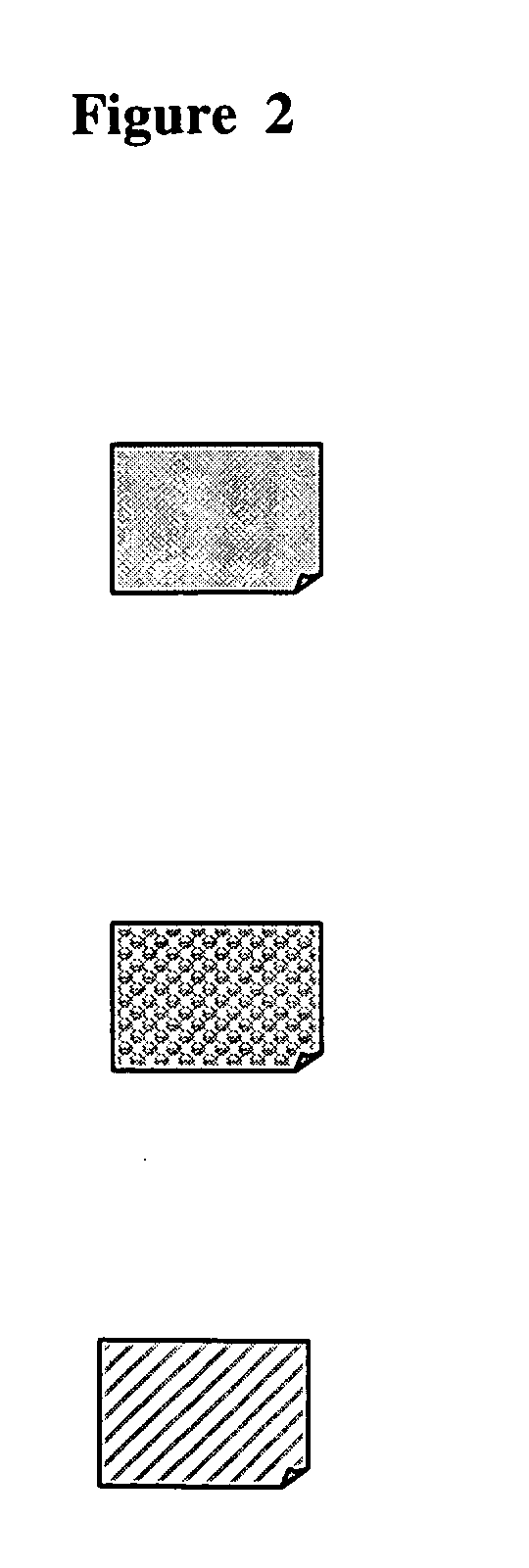
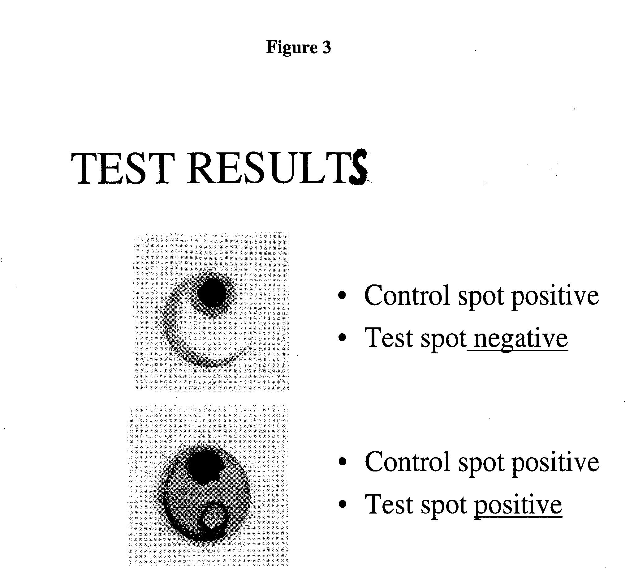
[0035] Yet another embodiment discloses a field test method for the detection of antibodies to S. neurona in equine serum or blood. A strip of nitrocellulose membrane is sandwiched between a lower absorbent layer of filter paper and a top layer of plastic foam in which a port or cutout is die-cut for the introduction of samples, reagents and wash buffers. A small sample of protein antibodies such as for instance, goat anti-mouse IgG, known to react nonspecifically with Protein A of the colloidal gold-Protein A conjugate, to yield a visible colored compound, is placed at a distinct spot on the nitrocellulose membrane assembly described. Excess IgG is washed off the nitrocellulose membrane with a suitable buffer. This serves as a control spot. A second small sample of the vaccine against S. neurona is also placed at another spot adjacent the IgG spot. This serves as the test spot. A serum or blood sample from a horse or other such animal, suspected of containing antibodies to S. neurona is obtained and contacted with the nitrocellulose membrane assembly with immobilized vaccine and the IgG. Unbound antibodies and excess serum are washed off with the buffer. The nitrocellulose layer is further contacted with a solution of colloidal gold conjugated with Protein A or Protein G to form a colored compound with the antigen-antibody complex and with the IgG. The intensity of the color of the compound at the test spot is determined as a indication of the concentration of the antibodies present in the body fluid of the horse or other animal. A serum sample from a healthy horse with no S. neurona induced antibodies serves as a negative control. The control test with IgG, and other tests with known samples for positive and negative results, establish the validity of the test and helps minimize false positives and false negatives.
[0046] Another embodiment provides a simple membrane-capturing assay for the presence of antibodies to a specific pathogen.

Problems solved by technology

Diseases caused by protozoal parasites are one of the most difficult for medical science to understand and control effectively.
Despite decades of research, significant understanding, diagnosis and treatment of the diseases have proved elusive.
Vaccines so far have been used as preventive agents but have not been used in diagnostic tests.
These methods are again, tedious, time consuming, require expert knowledge and training in immunochemical methods of analysis, such as the use of enzymes, radioisotopes or immuno-fluorescent markers as labels, washing off excess label and detecting and / or measuring the antibody-antigen complex formed.
Diagnostic testing in live animals poses many practical problems and does not always yield reliable results.
They also require several days to perform and are expensive.
The cost of wide testing to detect such cases can be prohibitive.
Furthermore, some of these tests are not highly specific to the pathogen and may yield false negatives or false positives.
“Helpful Tips” The Horse Report, Vol. 21 (1), University of California, Davis, January 2003, reports that current treatment of EPM using an anti-protozoal drug and the previous mode of treatment using a different anti-protozoal drug in combination with sulfonamide antimicrobial such as sulfadiazine with or without trimethoprim, are not very effective but seem to merely depress the parasite's viability such that the body's own immune system will have a chance to take over the control of the infection.
There are, however, severe adverse effects noted including deaths due to enterocolitis while administering the recommended dosages.
This method also requires laboratory equipment and trained personnel and takes many days before results can be obtained.
This assay uses a Western blot identification technique which requires long hours of incubation, electrophoresis, laboratory equipment and trained personnel and cannot be used in the field.
Similarly, many diseases caused by viruses and protozoal parasites have been particularly difficult to detect and diagnose early enough for timely treatment or require lengthy, time-consuming, expensive laboratory procedures.
There is no simple, quick field test for the early detection of any of these diseases.
Of particular interest for the equine industry and for veterinarians is that there is no fast and simple diagnostic test currently available which can be used in the field by non-technical personnel for the detection of S. neurona antibodies in the body fluids of horses and ungulates.
If the diagnosis is delayed, or worse, if there is a misdiagnosis and the horse is not treated in a timely manner, the treatment which is highly expensive and labor intensive, may have to be prolonged and even with that, there may be permanent neurological damage and eventual death.

Method used


Image

  • Novel field testing method and device for detection of equine protozoal myeloencephalitis
  • Novel field testing method and device for detection of equine protozoal myeloencephalitis
  • Novel field testing method and device for detection of equine protozoal myeloencephalitis

Examples


Embodiment Construction

[0057] The present invention provides a fast field test method for the diagnosis of many pathogens, specifically, protozoa, viruses, bacteria and nematodes and more specifically, S. neurona, which can be operated by someone with no special training or technical knowledge. The test method is a simple membrane-capturing assay that can be used in the field to detect the presence of S. neurona induced antibodies in body fluids such as blood, plasma, serum, urine, cerebrospinal fluid, tissue extracts and saliva, within about 3 to 5 minutes. One preferred embodiment of the invention is specifically directed to an immunologic method for the protozoa, S. neurona, by detecting antibodies present in the blood or serum of the infected animal, using a commercially available vaccine as the antigen therefor.

[0058] The present invention also provides a test device that utilizes antigens from killed S. neurona immobilized on a suitable support such as a nitrocellulose membrane. The antigens in the...


PUM


Abstract

The present invention discloses a quick, simple, qualitative and semi-quantitative immunologic method and device in a field test kit format for the detection and the stage of infection of certain protozoal, viral, bacterial, or nematodal pathogens in body fluids, using vaccines specific to such pathogens as antigens. The method comprises immobilizing an antigen specific to a selected protozoal, bacterial, viral or nematodal pathogen on a support membrane, obtaining a sample of a body fluid from a human or animal suspected of harboring the pathogen, thus containing antibodies thereto, contacting the antigen with the sample of body fluid and detecting the binding of the antigen with the antibodies by any suitable means. The device comprises a rigid member, an absorbent layer, a foam layer and a support member on which the antigen is immobilized, glued together to form a compact assembly. The field test kit comprises the device enclosed in a casing and vials containing additional reagents such as washing and dilution buffers, a chromogenic label for the detection of the antigen-antibody complex and positive and negative control solutions to verify the accuracy of the tests, instructions for use and the like. All the components are packaged in a small plastic container or box for easy transportation and use. While this invention is particularly adaptable to the detection of S. neurona using its vaccine as an antigen, other pathogens can also be detected by the method of this invention. These include but are not limited to Neospora hughesi, Neospora Canninum, Leishmania, Leptospira, West Nile virus, and other similar organisms.

Description

FIELD OF THE INVENTION [0001] The present invention relates to a novel and quick method for the qualitative and semi-quantitative diagnostic detection of infectious agents and pathogens such as protozoa, viruses, nematodes, bacterial and other microorganisms, including protozoal parasites such as Sarcocystis neurona, usually abbreviated S. neurona, which cause equine myeloencephalitis, commonly known as EPM, and other pathogens which cause diseases such as Neospora hughesi, Neospora Canninum, Leishmania, Leptospira, West Nile virus, and other similar organisms and the like and a field test device therefor. BACKGROUND OF THE INVENTION [0002] Diseases caused by protozoal parasites are one of the most difficult for medical science to understand and control effectively. Despite decades of research, significant understanding, diagnosis and treatment of the diseases have proved elusive. Vaccines have been developed against some of these pathogens and injected into the body of humans or an...

Claims


Application Information

Patent Timeline
IPC IPC(8): C12Q1/70G01N33/569G01N33/554G01N33/53
CPCG01N33/56905Y02A50/30
Inventor KRISTIPATI, RAMA SHARMASINGH, TEJINDERDUNNEBACKE, THELMA H.CALDWELL, ANTHONY
Owner KRISTIPATI RAMA SHARMA
Learn More