Treatment of glioblastoma with thymosin-alpha 1

a technology of thymosin and glioblastoma, which is applied in the direction of peptides, drug compositions, peptides, etc., can solve the problems of not always supporting the effectiveness of these compounds as a single agent on brain tumors, and the immune response to neoplastic cells is mainly ineffective in completely eradicating residual neoplastic cells, so as to prolong the survival of patients with glioblastomas, reduce tumor burden, and increase the production of

Inactive Publication Date: 2006-07-27
RHODE ISLAND HOSPITAL
View PDF4 Cites 0 Cited by
  • Summary
  • Abstract
  • Description
  • Claims
  • Application Information

AI Technical Summary

Benefits of technology

[0006] Glioblastomas are high-grade, malignant central nervous system (CNS) neoplasms that are nearly always fatal within 12 months of diagnosis. Recent studies showed that immunotherapy using pro-inflammatory cytokines such as IL-2 or IL-12 may prolong survival of patients with glioblastomas. Thymosin-α-1 (thymalfasin) is a thymic peptide that acts as an immune-modulator, increasing IL-2 production and T-cell proliferation. The present work demonstrated significantly reduced tumor burden and increased lympho-mononuclear inflammatory cell response in subjects treated with thymalfasin+BCNU relative to all other groups. In vitro experiments demonstrated that thymalfasin treatment had no direct effect on viability or mitochondrial function in cultured 9 L cells. However, thymalfasin treatment resulted in significantly increased levels of pro-apoptosis gene expression, including FasL, FasR and TNFα-IR (65.89%, 44.08% and 22.18%, respectively). In addition, thymalfasin treatment rendered the 9 L cells more sensitive to oxidative stress such that ordinarily non-lethal doses of H2O2 killed 30-50% of 9 L cells that had been treated with thymalfasin. Further studies revealed that thymalfasin enhances 9 L cell sensitivity to Granzyme B- (T cell) or BCNU-mediated killing. The results show that thymalfasin enhances chloroehtylnitrosurea-mediated eradication of glioblastoma in vivo, and that thymalfasin mediates its effects by activating pro-apoptosis mechanisms, rendering neoplastic cells more sensitive to oxidative stress and killing by Granzyme B (T cells) or chemotherapy.

Problems solved by technology

Glioblastomas cause death due to rapid, aggressive, and infiltrative growth in the brain.
In addition, the immune response to the neoplastic cells is mainly ineffective in completely eradicating residual neoplastic cells following resection and radiation therapy (Roth, 1999; Dix, 1999; Sablotzki, 2000).
Chloroethylnitrosureas have been utilized as a single treatment chemotherapy for many years on primary brain tumors; however, the historical statistics do not always appear to support the effectiveness of these compounds as a single agent on brain tumors (e.g., Aquafedda, et al.).

Method used


Image

  • Treatment of glioblastoma with thymosin-alpha 1
  • Treatment of glioblastoma with thymosin-alpha 1
  • Treatment of glioblastoma with thymosin-alpha 1

Examples


example 1

Thymosin-α1 Treatment of 9 L and 293 Tumor Cell Lines

[0025] Tumor cell lines: Rat 9 L glioblastoma cells and 293 human kidney cells were maintained in Dulbecco's Modified Eagle's Medium (DMEM) supplemented with 5% FCS, 2 mM L-glutamine, and 100 μM non-essential amino acids (Gibco-BRL, Grand Island, N.Y.). All cell lines were kept at 37° C. in a humidified atmosphere containing 5% CO2. The cell lines were obtained from the American Type Culture Collection and were certified as free of pathogens.

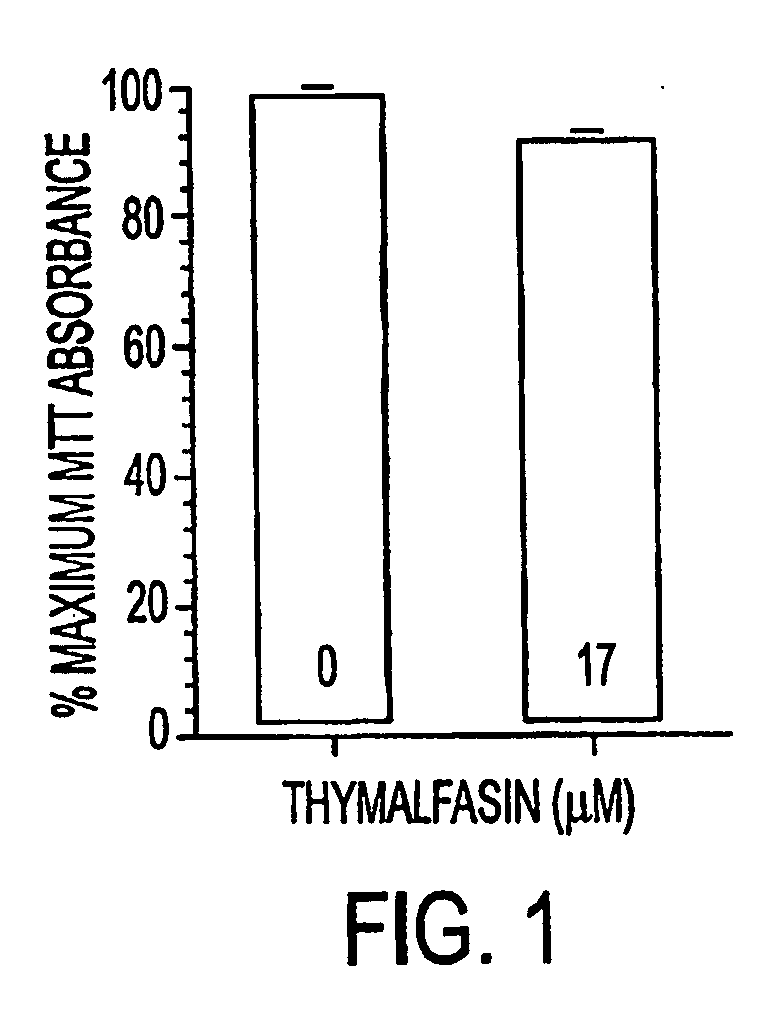
[0026] Thymosin alpha 1 treatment: For acute thymalfasin treatment, cells seeded in 96-well plates at a cell density of 20,000 cells / well were treated with 10−5 M thymalfasin for 24 hours. Cells treated chronically with thymalfasin were grown in flasks and treated every 24 hours with 10−5 M thymalfasin (added to fresh medium) for the duration of the treatment (3 days). In addition, for the determination of a dose-response curve to thymalfasin for 9 L cells, cells were treated with serially d...

example 2

Granzyme-Induced Apoptosis Studies

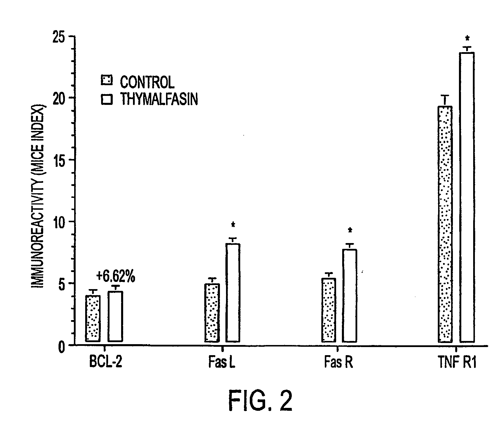
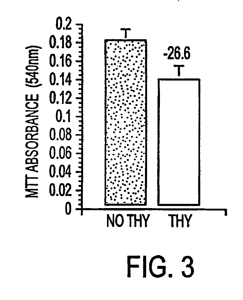
[0034] The MICE assay studies described above demonstrated thymalfasin-induced expression of TNF-R1, FasL and FasR in 9 L glioblastomas. In order to determine the differential susceptibility of 9 L glioblastoma cells treated (or not) with thymalfasin to cytotoxic T-lymphocyte (CTL) induced apoptosis, experimental assays were conducted in which 9 L cells were treated both acutely for 24 hours and chronically for 72 hours with thymalfasin, after which they were harvested, re-seeded into 96-well black plates (7.5×10⁵ cells / 75 μl / well), and exposed to 20,000 units / ml Streptolysin O (SLO) plus 100 ng recombinant Granzyme B (reaction volume 100 μl) for 1 or 3 hours at 37° C. The SLO was used in place of perforin to permeabilize the cells, and recombinant Granzyme B was used to standardize the assay. Control studies included parallel reactions in which SLO, Granzyme B, or both were omitted. Viable cell density was measured using the ATPlite assay (Packard...

example 3

Effect of Thymalfasin on Cell Viability of Primary Neuronal Cell Cultures

[0036] Primary cortical neuron cultures were studied to enable selection of thymalfasin doses that would not be toxic to non-neoplastic brain cells. Cell viability and mitochondrial function were measured using the Crystal violet (CV) and MTT assays since previous studies showed that CV and MTT absorbances increase linearly with cell density from 1×10⁴ to 5×10⁵ cells per well (de la Monte, 2001 & 2000).

[0037] Primary neuronal cortical cells treated in 96-well plates with serial dilutions of thymalfasin (final concentrations ranging from 3.3×10⁻⁵ M to 1×10⁻⁹ M) showed no decrease in cell viability at the experimental doses used. The highest concentration of thymalfasin used (3.3×10⁻⁷M) did show a 30% decrease in viability as seen through MTT assay. However, this dose is higher than established experimental and clinical dosages.


PUM

PropertyMeasurementUnit
pHaaaaaaaaaa
pHaaaaaaaaaa
volumeaaaaaaaaaa

Abstract

Thymosin-α1 is used as an adjuvant in combination with carmustine (BCNU) as an effective treatment for malignant glioblastoma.

Description

RELATED APPLICATION DATA [0001] This application claims the benefit of provisional application 60 / 337,149, filed Dec. 10, 2001.BACKGROUND OF THE INVENTION [0002] Glioblastoma is the most common primary CNS malignant neoplasm in adults, and accounts for nearly 75% of the cases. Although there has been steady progress in their treatment due to improvements in neuro-imaging, microsurgery and radiation, glioblastomas remain incurable (McDonald, 2001; Burton, 2000; Prados, 2000). The average life expectancy is less than one year from diagnosis, and the five-year survival rate following aggressive therapy including gross tumor resection is less than 10% (Burton, 2000; Nieder, 2000; Napolitano, 1999; Dazzi, 2000). Glioblastomas cause death due to rapid, aggressive, and infiltrative growth in the brain. The infiltrative growth pattern is responsible for the un-resectable nature of these tumors. Glioblastomas are also relatively resistant to radiation and chemotherapy, and therefore post-tre...

Claims


Application Information

Patent Timeline
Patent Type & Authority Applications(United States)
IPC IPC(8): A61K38/17A61K31/175A61K38/00A61K31/17A61K38/22A61K45/06A61P35/00
CPCA61K31/17A61K31/175A61K45/06A61K2300/00A61K38/2292A61P35/00A61K38/16A61K31/64
Inventor WANDS, JACK R.DE LA MONTE, SUZANNE
Owner RHODE ISLAND HOSPITAL
Why Eureka
Try Eureka