
Method of administering split doses of a vascular targeting agent

a vascular targeting agent and split dose technology, applied in the field of split doses of vascular targeting agents, can solve the problems of avalanche of ischaemic tumor cell death, unable to effectively treat patients, and unable to achieve effective treatment, so as to achieve less effective effect and more cell killing

Inactive Publication Date: 2006-06-22
OXIGENE
View PDF1 Cites 0 Cited by
  • Summary
  • Abstract
  • Description
  • Claims
  • Application Information

AI Technical Summary

Benefits of technology

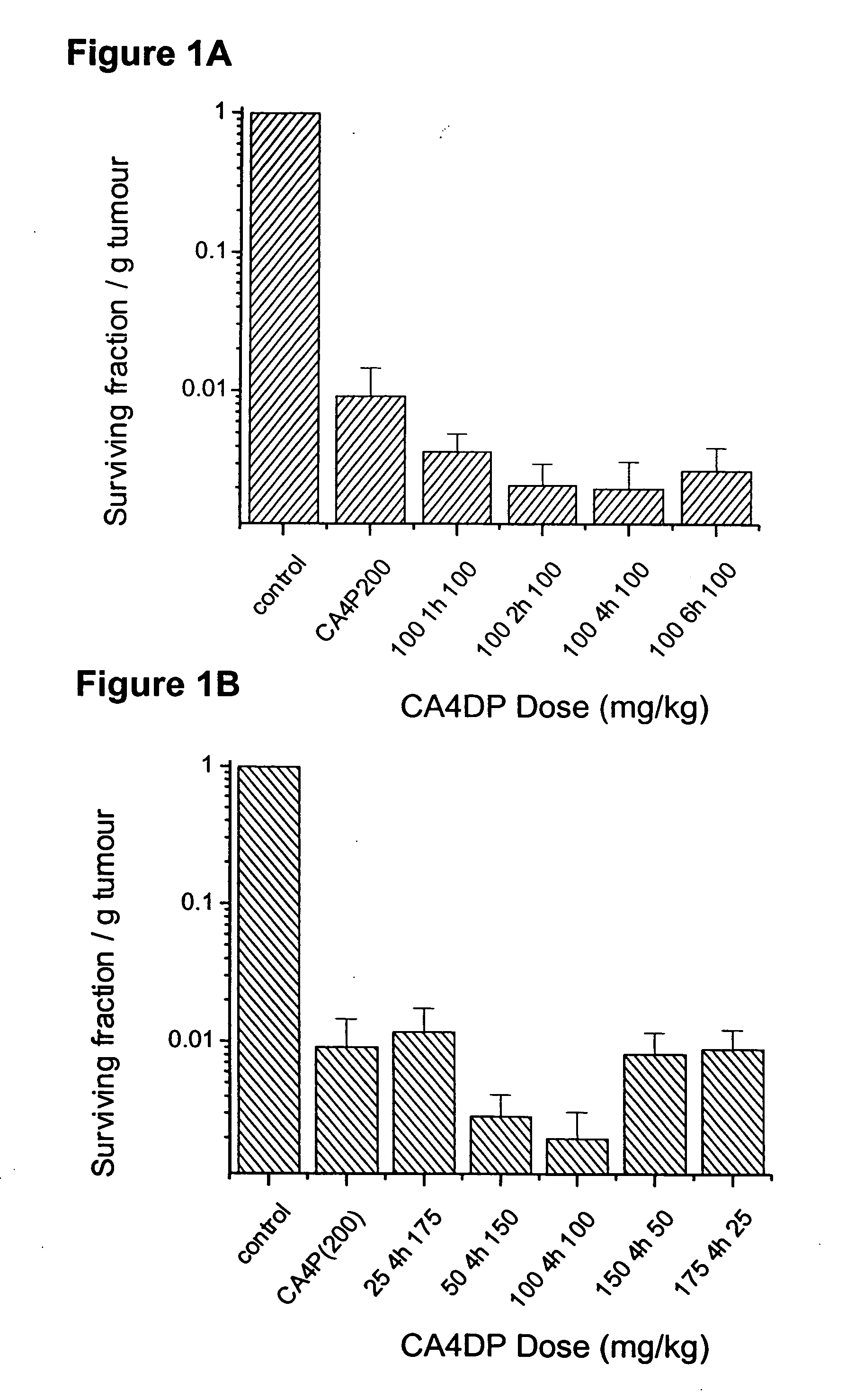
[0036] To compare the anti-tumor effect of single and divided VTA dose scheduling, the survival of tumor cells isolated from a treated CaNT tumor-bearing animals was assessed by an in vitro clonogenic (colony-forming) assay, as described previously (Chaplin et al., Anticancer Research, 1999, 19: 189-196). Tumors were excised 18-24 h after CA4P injection. Two tumors were combined for each data point, weighed, minced with scissors and then disaggregated for 1 hour at 37° C. in an enzyme cocktail of 1 mg/ml pronase, 0.5 mg/ml DNase and 0.5 mg/ml collagenase. Following digestion, samples were passed through a 25G needle and a 35 μm filter to obtain a single cell suspension. Haemocytometer counts of trypan-blue excluding cells were made and known numbers of viable cells added to a feeder layer of heavily irradiated V79-379A Chinese hamster cells. After 7-10 days incubation, colonies were fixed, stained with methylene blue and counted. The data were calculated as surviving fraction per gram of tumor, which is a product of relative surviving fraction and relative cell yield per gram...

Problems solved by technology

Cancer is a leading cause of death in the industrialized world and despite years of research many types of cancer lack an effective therapeutic treatment.
This is especially true for cancers that are characterized by the presence of large, solid tumors since it is difficult to deliver an effective dose of a chemotherapeutic agent to the interior of a large tumor mass with a degree of selectivity.
Moreover, due to the genetic instability of tumor cells, a tumor tissue can rapidly acquire resistance to standard therapeutic regimens.
The disruption in the function of a single tumor blood vessel can result in an avalanche of ischaemic tumor cell death and necrosis of thousands of cancer cells which depend on it for blood supply.
A single dose of VTA can cause a rapid and selective shutdown of the tumor neovasculature within a period of minutes to hours, leading eventually to tumor necrosis by induction of hypoxia and nutrient depletion.
Other agents have been known to disrupt tumor vasculature but differ in that they also manifest substantial normal tissue toxicity at their maximum tolerated dose.

Method used


Image

  • Method of administering split doses of a vascular targeting agent
  • Method of administering split doses of a vascular targeting agent
  • Method of administering split doses of a vascular targeting agent

Examples


example 1

Effect of Split VTA Dosing on Tumor Cell Survival

Methods and Materials

[0036] To compare the anti-tumor effect of single and divided VTA dose scheduling, the survival of tumor cells isolated from a treated CaNT tumor-bearing animals was assessed by an in vitro clonogenic (colony-forming) assay, as described previously (Chaplin et al., Anticancer Research, 1999, 19: 189-196). Tumors were excised 18-24 h after CA4P injection. Two tumors were combined for each data point, weighed, minced with scissors and then disaggregated for 1 hour at 37° C. in an enzyme cocktail of 1 mg / ml pronase, 0.5 mg / ml DNase and 0.5 mg / ml collagenase. Following digestion, samples were passed through a 25G needle and a 35 μm filter to obtain a single cell suspension. Haemocytometer counts of trypan-blue excluding cells were made and known numbers of viable cells added to a feeder layer of heavily irradiated V79-379A Chinese hamster cells. After 7-10 days incubation, colonies were fixed, stained with methylen...

example 2

Effect of Split VTA Dosing on Tumor Growth Delay

Material and Methods

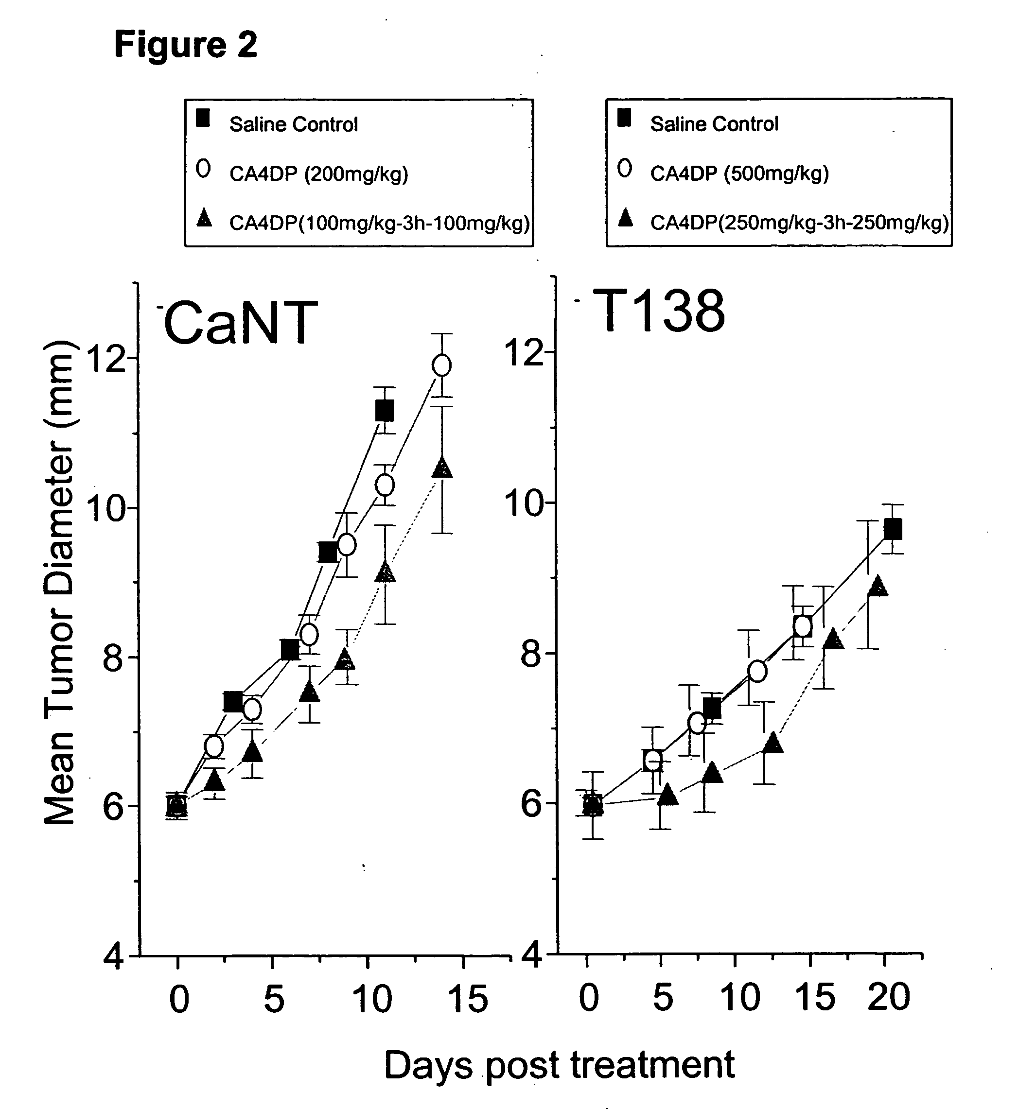
[0038] The anti-tumor effect of split dosing therapy was also assessed by an analysis of tumor growth delay in both CaNT and T138 tumor-bearing mice. For experiments involving the CaNT, dose groups comprised 5 to 6 animals, whereas for the T138, 10 to 15 animals were used. Following initial drug treatment, tumor growth was determined by measuring the diameter of each tumor in 3 orthogonal orientations two or three days each week.

Results

[0039] The results of split dose therapy on tumor growth delay are illustrated in FIG. 2. As in Example 1, equal split dose therapy resulted in an enhanced tumor response over a single dose of CA4DP. This was apparent in the CaNT mice, where a single 200 mg / kg dose of CA4DP was compared with two equal doses of 100 mg / kg separated by a time interval of 3 hours. A single dose of CA4DP dose did not delay growth relative to control, while split dose therapy delayed tumor growth for ...

example 3

Effect of Split VTA Dosing on the Vascular Perfusion of Tumors

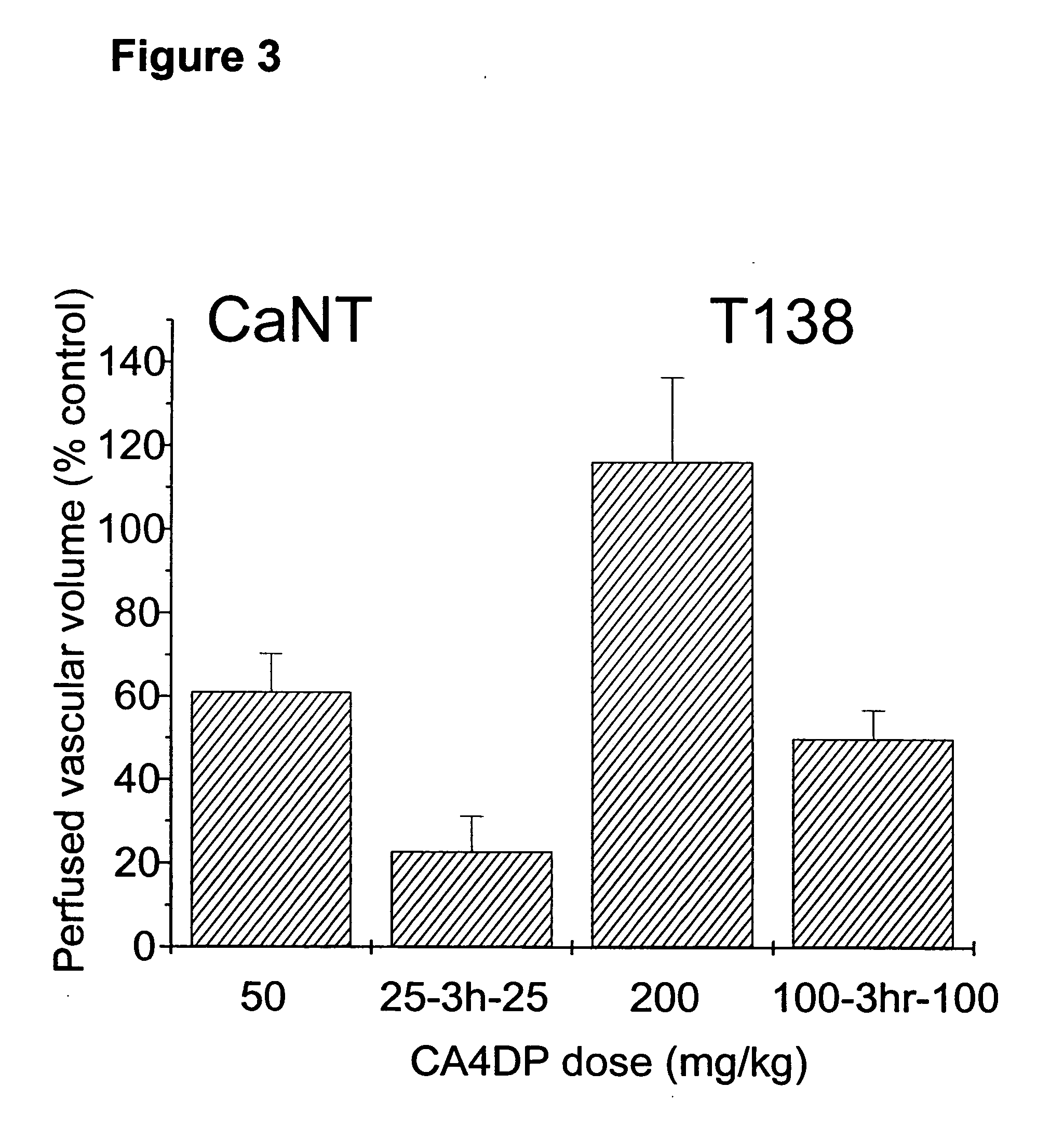
[0040] The effect of VTA dose splitting on the tumor vascular response was also investigated in both CaNT and T138 tumor types, by measuring the functional vascular volume at 24 hours following treatment with single or split doses of CA4DP. Functional vascular volume is defined as the volume of tumor tissue perfused by tumor blood vessels, and is measured using the fluorescent DNA-binding dye Hoechst 33342 (Sigma-Aldrich Company Ltd., Dorset, UK) (Smith et al, British Journal of Cancer, 1988, 57: 247-253). The dye was dissolved in 0.9% saline at 6.25 mg / ml and injected i.v. at a dose of 10 mg / kg at 24 hours post-treatment. Tumors were excised and frozen 1 minute later. Sections were cut at three levels and observed under UV illumination. Functional vessels were identified by the fluorescent outline of perivascular tissue and vascular volumes were determined using a random point scoring system based on that described by C...


PUM

PropertyMeasurementUnit
Timeaaaaaaaaaa
Timeaaaaaaaaaa
Timeaaaaaaaaaa

Abstract

The present invention is directed to the use of vascular targeting agents or pharmaceutically acceptable salts thereof for administration in divided doses to a warm-blooded animal, such as a human. Also disclosed is a medicament comprising two or more fraction of doses of a vascular targeting agent, or a pharmaceutically acceptable salt thereof, which together add up to a total daily dose, or administration in divided doses for use in a method of treating a human or warm-blooded animal. A kit comprising two or more fractions of doses of a vascular targeting agent or a pharmaceutically acceptable salt thereof, which together add up to a total daily dose, for administration in divided doses is also disclosed.

Description

RELATED APPLICATIONS [0001] This is a continuation of U.S. application Ser. No. 10 / 265,820, filed Oct. 7, 2002. The entire contents of the above identified application is incorporated herein by reference.FIELD OF THE INVENTION [0002] The present invention provides a method of administering Vascular Targeting Agents (“VTAs”) to treat diseases associated with malignant neovascularization. BACKGROUND OF THE INVENTION [0003] Cancer is a leading cause of death in the industrialized world and despite years of research many types of cancer lack an effective therapeutic treatment. This is especially true for cancers that are characterized by the presence of large, solid tumors since it is difficult to deliver an effective dose of a chemotherapeutic agent to the interior of a large tumor mass with a degree of selectivity. Moreover, due to the genetic instability of tumor cells, a tumor tissue can rapidly acquire resistance to standard therapeutic regimens. [0004] In order to develop into a l...

Claims


Application Information

Patent Timeline
IPC IPC(8): A01N31/14A61K9/22A61K31/075
CPCA61K31/075
Inventor CHAPLIN, DAVID J.HILL, SALLY
Owner OXIGENE
Learn More