Compositions for detecting hyaluronidase activity in situ and methods of utilizing same
a technology of hyaluronidase activity and compositions, applied in the field of compositions and methods for detecting hyaluronidase activity in situ, can solve the problem that none of these methods can be applied in-situ for non-invasive imaging
- Summary
- Abstract
- Description
- Claims
- Application Information
AI Technical Summary
Benefits of technology
Problems solved by technology
Method used
Image
Examples
example 1
Detection of Hyaluronidase Activity by MRI using HA-GdDTPA-Beads
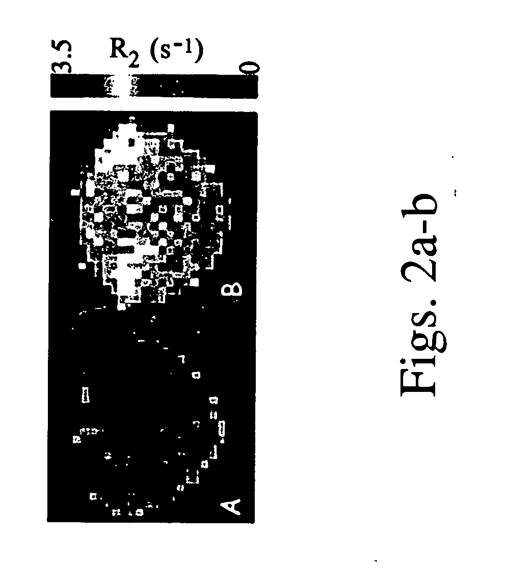
[0115] Avidin-agarose beads, were covalently linked via the avidin moiety to HA-GdDTPA (FIG. 1), in order to develop a contrast material for non-invasive imaging of hyaluronidase activity by MRI. Suspension of HA-GdDTPA-beads (1 mg / ml) was used to test the ability of MRI to detect hyal-dependent changes in the R2 relaxation rate of water.
[0116] As shown in FIGS. 2a-b, a significant increase in R2 was measured for beads supplemented with the commercial hyaluronidase relative to control HA-GdDTPA-beads suspended in pure DDW (1 tail t-test unpaired p=0.0047).
example 2
Expression of Hyal-1 and Hyal-2 in Ovarian Carcinoma Cells
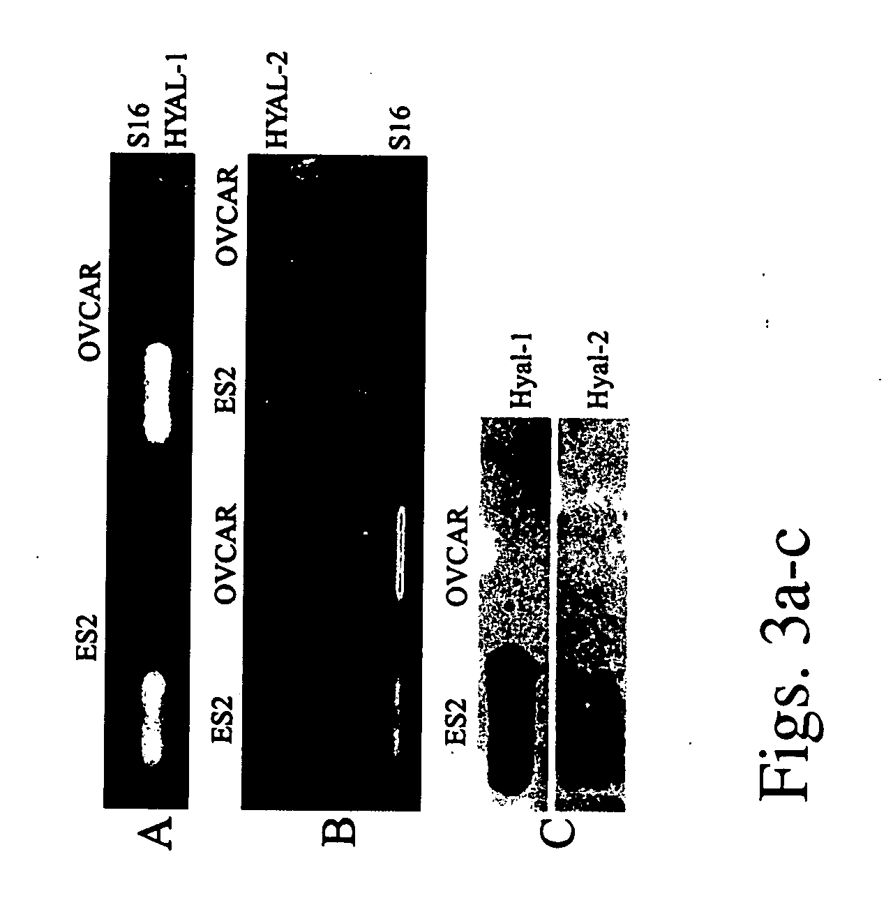
[0117] The expression of hyaluronidase 1 and 2 was measured in two human epithelial ovarian carcinoma cell lines: OVCAR-3 and ES-2. Semi-quantitative Reverse Transcription PCR showed no significant difference in the mRNA levels of HYAL-1 or HYAL-2 between those 2 cell lines (FIGS. 3a-b).
[0118] In contrast with the similarity in mRNA expression, the amount of hyaluronidase released to the culture medium was significantly different. The conditioned medium of the cells was analyzed by Western blot analysis using anti-Hyal-1 and anti-Hyal-2 antibodies. A large difference was detected in the amount of hyaluronidase secreted by OVCAR-3 and ES-2 cells (FIG. 3c). Medium conditioned by ES-2 cells demonstrated high levels of both Hyal-1 and Hyal-2, while in medium conditioned by OVCAR-3 cells the proteins were un-detectable. Although ES-2 cells exhibit higher rate of proliferation in 10% FCS, they showed decreased cell survival in se...
example 3
Secretion of Active Hyaluronidase by Human Epithelial Ovarian Carcinoma Cells
[0119] Particle exclusion assay was used to evaluate the activity of hyaluronidase secreted by ovarian carcinoma cells (FIGS. 4a-e). The chondrocytes used in this assay were surrounded by a thick pericellular coat comprised of high molecular weight HA, which excludes red blood cells. Hence, red blood cells which are added to the culture plate, surround the HA layer, delineating it and thereby allowing its visualization (FIG. 4a). Upon addition of hyaluronidase, the high molecular weight HA is degraded, allowing the red blood cells to approach the chondrocyte membrane (FIG. 4b).
[0120] The activity of hyaluronidase in medium conditioned by OVCAR-3 and ES-2 ovarian carcinoma cells was examined by addition of this medium to the chondrocytes (FIG. 4c-d). Conditioned medium from ES-2 cells degraded the HA layer and showed significantly higher activity of hyaluronidase relative to conditioned medium from OVCAR-3...
PUM
| Property | Measurement | Unit |
|---|---|---|
| molecular weight | aaaaa | aaaaa |
| atomic number | aaaaa | aaaaa |
| atomic number | aaaaa | aaaaa |
Abstract
Description
Claims
Application Information
 - R&D
- Intellectual Property
- Life Sciences
- Materials
- Tech Scout
- Unparalleled Data Quality
- Higher Quality Content
- 60% Fewer Hallucinations
Browse by: Latest US Patents, China's latest patents, Technical Efficacy Thesaurus, Application Domain, Technology Topic, Popular Technical Reports.
© 2025 PatSnap. All rights reserved.Legal|Privacy policy|Modern Slavery Act Transparency Statement|Sitemap|About US| Contact US: help@patsnap.com


