Compositions and methods for phage display of polypeptides
a polypeptide and phage technology, applied in the field of new phage display vectors, can solve the problems of loss of infectivity, reducing or even preventing successful enrichment of polypeptides, and the artisan seeking to employ phage display methods, so as to increase the proportion of rgps
- Summary
- Abstract
- Description
- Claims
- Application Information
AI Technical Summary
Benefits of technology
Problems solved by technology
Method used
Image
Examples
example 1
Materials
[0082] The following materials are used in the following methods: E. coli strains (JM109, CJ236, DH12S), 3.0 μM streptavidin magnetic latex (M280,Dynal Biotech), thrombin (Novagen), 2xYT, 10× thrombin digestion buffer (200 mM Tris-HCl, pH 8.4, 1.5M NaCl, 25 mM CaCl2), PEG / NaCl solution (30% PEG6000, 3M NaCl), panning buffer (Tris 40 mM, 150 mM NaCl, 20 mg / ml BSA, 0.1% Tween20, pH 7.5), elution buffer (0.1M glycine buffer pH 2.0), Roche complete protease inhibitor cocktail EDTA-free, goat anti-murine K-biotin (Southern Biotech), sheep anti-geneVIII-biotin (Seramune, biotinylated at Biosite as described in Example 10 of U.S. Pat. No. 6,057,098), reduced ampletamine-BSA-Biotin (“rMET-BSA-Biotin,” prepared as described in Example 4 of U.S. Patent Publication 20030162249), mutagenic oligonucleotide #1823 5′Phos-CAGACAAGCACTAGTAGAA CCACGCGGAACCAGAGAACCGCCACCAGTGCCACCGCCAGAATCCCTGGGCAC.
Example 2
Selection of Proteases and Proteolytic Cleavage Sites
[0083] The selection of an app...
example 2
Display Vector Preparation
[0085] A type 3 phage-display vector displaying a murine anti-rMET Fab, Fdcox24.6, was chosen as a model system. This vector has the carboxy-terminus of the heavy chain domain CH1 fused to the 7th amino acid of mature gene III (Dower, EP 0527839, the contents of which are incorporated by reference herein in their entirety, including all tables, figures, and claims). DNA encoding a peptide linker (GGGTGGGS) followed by a thrombin recognition site (LVPRGS) was introduced into Fdcox24.6 DNA between the carboxy terminus of the heavy chain and the 7th amino acid of mature gene III by mutagenesis (Kunkel, U.S. Pat. No. 4,873,192, the contents of which are incorporated by reference herein in their entirety, including all tables, figures, and claims) using Fdcox24.6 uracil template made in CJ236 and oligonucleotide #1823. The completed mutagenesis reaction was electroporated into E. coli strain DH12S (Invitrogen). Incorporation of the correct sequence into Fdtet24...
example 3
Analysis of Bacteriophage Infectivity
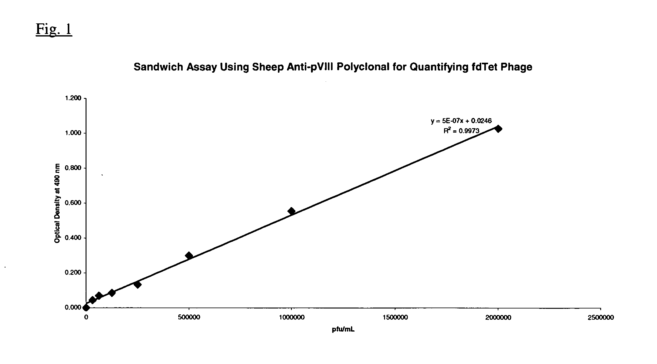
[0086] Fdcox24.6T was grown overnight in 50 ml of 2xYT (20 μg / ml tet) at 30° C., with shaking. This culture was used at a 1 / 100 dilution to inoculate 500 ml of 2xYT (20 μg / ml tet). After overnight growth at 30° C., with shaking, the bacterial cells were pelleted by centrifugation and the clarified supernatant containing Fdcox24.6T phage (presenting murine Fab) was harvested by centrifugation. The Fdcox24.6T phage was precipitated from 140 ml of clarified supernatant by the addition of ⅕ volume of a PEG / NaCl solution. After incubation at 4° C. for one hour, the phage were pelleted by centrifugation and thoroughly resuspended in 1.0 ml of PBS. The sample was centrifuged to pellet residual bacterial debris and the phage containing supernatant transferred to a fresh tube and stored at 4° C. A titer was determined by the following ELISA assay since the low infectivity of the fusion phage does not produce visible plaques when plated on an appropriate ...
PUM
| Property | Measurement | Unit |
|---|---|---|
| pH | aaaaa | aaaaa |
| pH | aaaaa | aaaaa |
| pH | aaaaa | aaaaa |
Abstract
Description
Claims
Application Information
 - R&D
- Intellectual Property
- Life Sciences
- Materials
- Tech Scout
- Unparalleled Data Quality
- Higher Quality Content
- 60% Fewer Hallucinations
Browse by: Latest US Patents, China's latest patents, Technical Efficacy Thesaurus, Application Domain, Technology Topic, Popular Technical Reports.
© 2025 PatSnap. All rights reserved.Legal|Privacy policy|Modern Slavery Act Transparency Statement|Sitemap|About US| Contact US: help@patsnap.com


