
Chimeric metabotropic glutamate receptors and uses thereof

Inactive Publication Date: 2005-08-25
ASTRAZENECA AB
View PDF18 Cites 8 Cited by
  • Summary
  • Abstract
  • Description
  • Claims
  • Application Information

AI Technical Summary

Benefits of technology

[0030] As indicated, an advantageous use of the constructs and methods of the present invention is to screen for compounds which modulate metabotropic glutamate receptor activity and to use such compounds to aid in the treatment of neurological diseases or disorders.
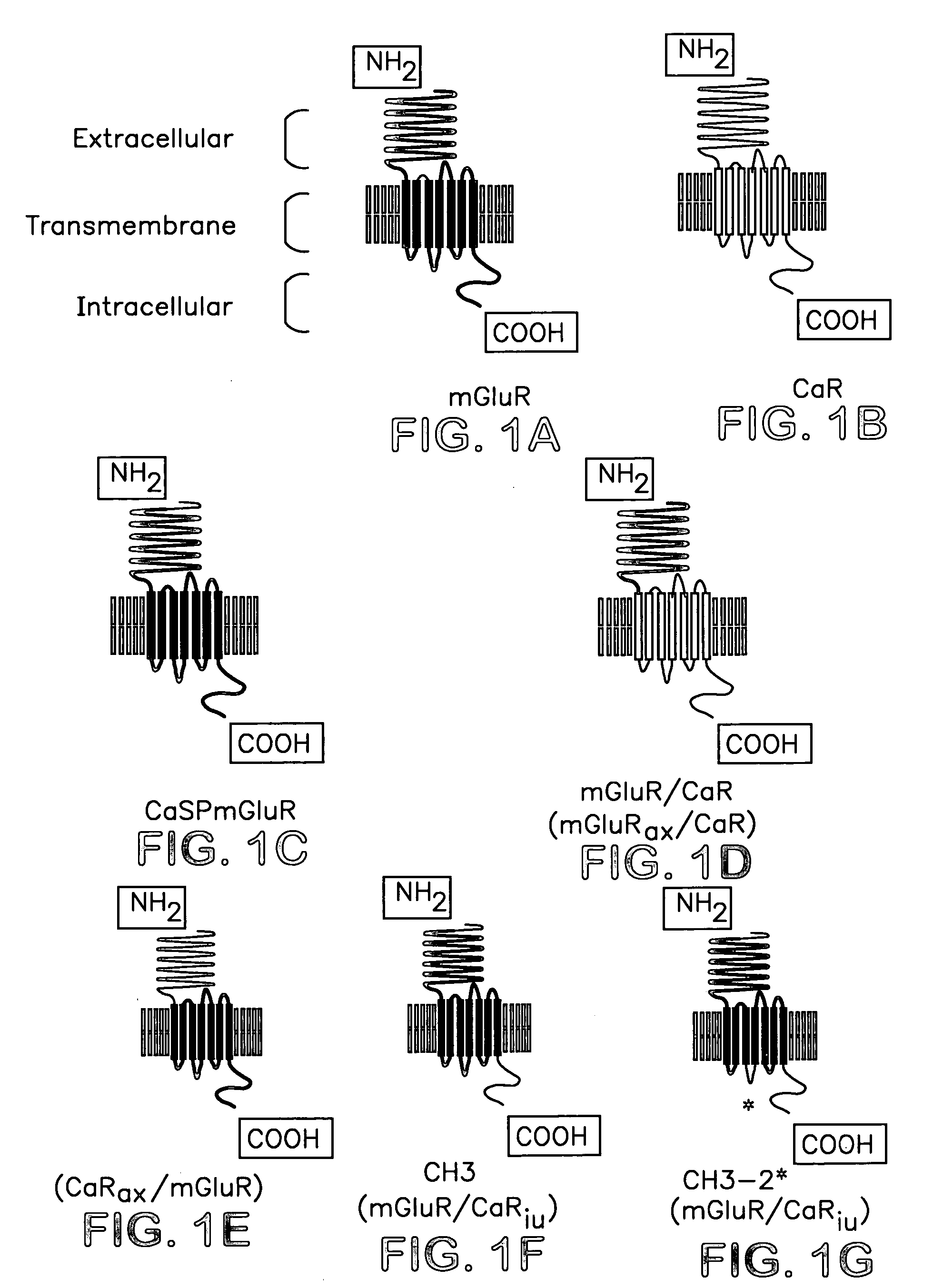
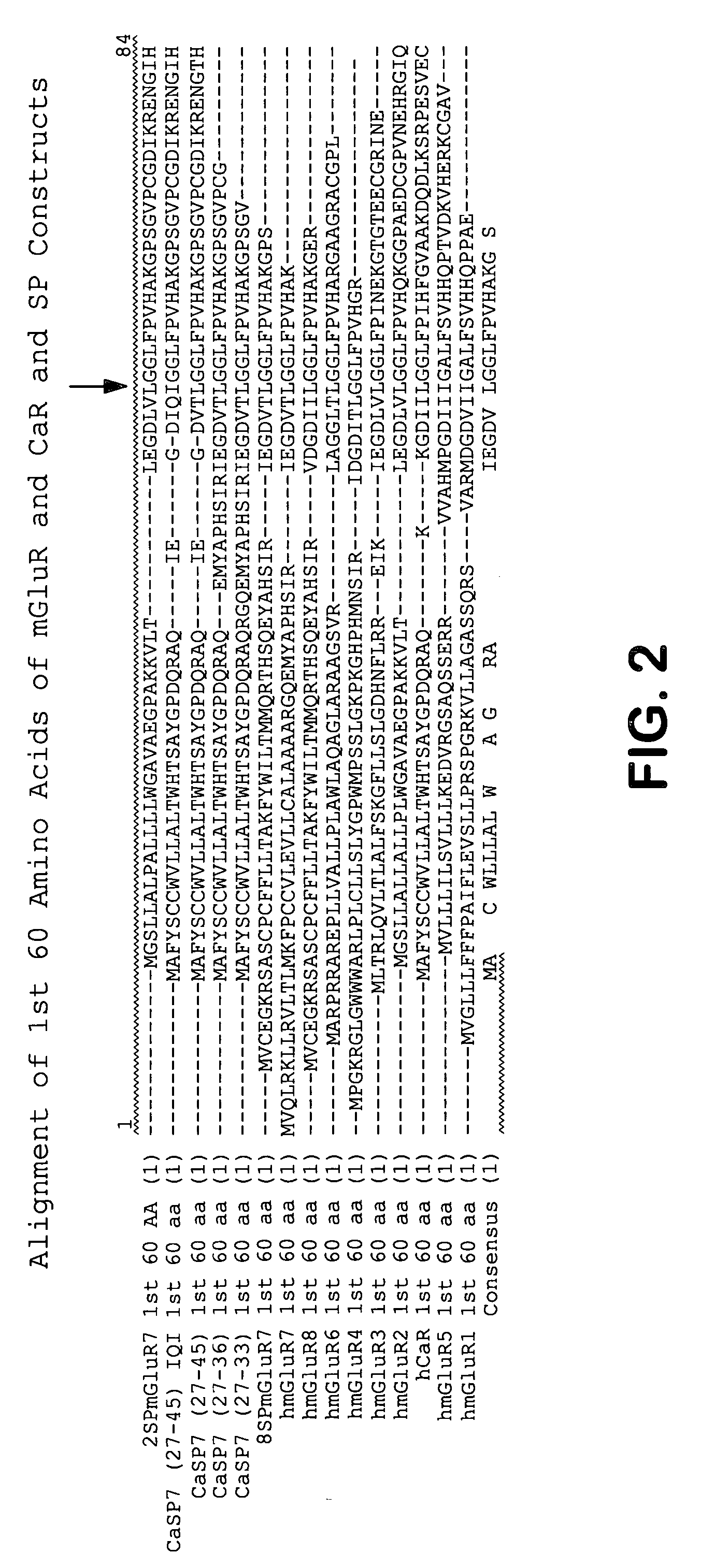
[0031] As described in the Background of the Invention above, metabotropic glutamate receptors (mGluR) and calcium receptors (CaR) have similar structures. Both types of receptors have an extracellular domain (ECD), a seven transmembrane domain (7TMD) and an intracellular cytoplasmic tail (CT). The present chimeric receptors include an extracellular domain that is the same as or has a high level of sequence identity to an mGluR and a signal peptide (SP) that is from a non-native source (i.e., not naturally associated with the mGluR from which the chimeric receptor mGluR sequence is obtained or derived), e.g., is from a CaR or a different mGluR or has a high level of sequence identity to a SP from such different source. Changing the signal peptide in this manner can provide increased expression of an mGluR and / or provide an epitope that makes assaying and / or visualizing the presence of such receptors more convenient than native receptors. For example, an antibody can be used that recognizes a fragment derived from the non-native signal peptide or the junction between the non-native signal peptide and the mGluR sequence.
[0039] The use of mGluRs for screening for mGluR active compounds has been complicated by a number of factors including a rapid desensitization of the receptor upon ligand binding / activation and difficulties in stably expressing the receptors in recombinant vertebrate cells (see, for example, FIG. 6B). Certain of the chimeric receptors of the present invention can be utilized to overcome these technical difficulties and provide much improved screening methods by utilizing the more robust aspects of calcium receptors. For example, by coupling the 7TMD and the CT of the CaR with the extracellular domain of an mGluR, or the CT of the CaR to the ECD and 7TMD of the mGluR, the mGluR extracellular domain has the benefit of the Gq coupling property of a CaR as well as the improved property of a lack of rapid densensitization (see, for example, FIG. 6C). Thus, such a chimeric receptor has the ligand binding and activation properties similar to those of a native mGluR but having the improved second messenger coupling similar to a CaR. Therefore, the chimeric receptor simplifies and enables efficient, practical, and reproducible functional screens to identify mGluR active molecules.

Problems solved by technology

A further level of complexity may be introduced by multiple interactions between mGluR expressing neurons in a given brain region.
Evidence for physiological effects of Group II mGluR activation at the postsynaptic level is limited.
This limits the range of pharmacological properties and potential therapeutic utilities of such compounds.
Furthermore, the range of pharmacological specificities associated with these mGluR- active molecules does not allow for complete discrimination between different subtypes of metabotropic glutamate receptors (Pin et al., Neuropharmacology 34:1, 1995 and Knopfel et al., J. Med. Chem. (1995) 38:1418).
Indeed, no mGluR-active molecules are presently under clinical development.
Gabellini et al., (Neurochem. Int. 24:533, 1994) also noted difficulties with mGluR1 expression in HEK 293 cells and it is possible that some of these difficulties may be due to desensitization characteristics of these receptors.
Furthermore, screening methodologies useful for identification of compounds active at Class I mGluRs are not readily amenable to identification of compounds active at class II and III mGluRs and vice versa due to the differences in second messenger coupling.
Finally, mGluRs have been noted to rapidly desensitize upon agonist stimulation which may adversely affect the viability of cell lines expressing these receptors and makes the use of native mGluRs for screening difficult.
While such experiments indicate that the third intracellular loop plays an important role in determining the specificity of G protein coupling in these related receptors, they have failed to identify any specific amino acid sequence motif which is responsible.
Thus, sequence conservation between CaRs and mGluRs appears to be consistent mostly with conservation of structural domains involved in ligand binding and G-protein coupling and does not provide evidence for specific sequence motifs within intracellular regions predictive of G-protein coupling specificity.

Method used


Image

  • Chimeric metabotropic glutamate receptors and uses thereof
  • Chimeric metabotropic glutamate receptors and uses thereof
  • Chimeric metabotropic glutamate receptors and uses thereof

Examples


example 1

Functional Expression in Oocytes

[0278] Oocytes suitable for injection were obtained from adult female Xenopus laevis toads using procedures described in C. J. Marcus-Sekura and M. J. M. Hitchcock, Methods in Enzymology, Vol. 152 (1987). Pieces of ovarian lobe were incubated for 30 minutes in Ca2+-free Modified Barths Saline (MBS) containing 1.5 mg / ml collagenase type IA (Worthington). Subsequently, 5 ng of RNA transcript prepared as described below, were injected into each oocyte. Following injection, oocytes were incubated at 16° C. in MBS containing 0.5 mM CaCl2 for 2-7 days prior to electrophysiological examination.

[0279] RNA transcripts encoding the chimeric receptors, or the GIRK subunits described below, were produced by enzymatic transcription from plasmid templates using T7 polymerase supplied with the mMessage mMachine ™(Ambion). Each plasmid was treated with a restriction enzyme to make a single cut distal to the 3′ end of the cDNA insert to linearize the template. This ...

example 2

Transfection and Growth of HEK293 Cells to Express Chimeric Receptors

[0284] A. Lipofectamine™ 2000 Transfections

[0285] Human embryonic kidney cells (HEK293, ATCC, CRL 1573) were maintained and propagated in culture in a routine manner. 20×106 cells were plated in T150 cm2 cell culture flasks in Dulbecco's Modified Eagle's Medium (DMEM from Gibco Life Technologies) containing 10 % fetal bovine serum (FBS from Hyclone Laboratories) to attain a monolayer of 95% confluence in 48-hours. To prepare plasmid DNA for transfection, the cDNA was precipitated with ethanol, rinsed and resuspended in sterile water at a concentration of 1 μg / ul. Sixty-three μg of cDNA was incubated with 197.5 μl of the liposome formulation Lipofectamine™ 2000 transfection reagent (Invitrogen) for 20 minutes in 4 ml serum-free Opti-MEM™ (Gibco Life Technologies) at room temperature allowing for the formation of the DNA-Cationic lipid complex. Post incubation, the 4 ml of complex was added to 40 ml of Opti-MEM™ in...

example 3

Measuring Changes in Intracellular Calcium Caused by Activation of Chimeric Receptors by the Fura Assay

[0288] Measurements of intracellular calcium release in response to increases in extracellular calcium is quantitated using the Fura assay (Parks et al. 1989). Stably transfected cells containing chimeric receptors are loaded with 2 μM fura-2 acetoxymethylester by incubation for 20-30 minutes at 37° C. in SPF-PCB (126 mM NaCl, 5 mM KCl, 1 mM MgCl2, 20 mM HEPES, pH 7.4), containing 1.25 mM CaCl2, 1 mg / ml glucose, 0.5% BSA1. The cells are then washed 1 to 2 times in SPF-PCB containing 0.5 mM CaCl2, 0.5% BSA and resuspended to a density of 4 to 5 million cells / ml and kept at 22° C. in a plastic beaker. For recording fluorescent signals, the cells are diluted fivefold into a quartz cuvette with BSA-free 37° C. SPF-PCB to achieve a final BSA concentration of 0.1% (1.2 ml of 37° C. BSA-free SPF-PCB+0.3 ml cell suspension). Measurements of fluorescence are performed at 37° C. with consta...


PUM

PropertyMeasurementUnit
Fractionaaaaaaaaaa
Nucleic acid sequenceaaaaaaaaaa

Abstract

The present invention provides chimeric receptors that include an extracellular domain from a metabotropic glutamate receptor and a non-native signal peptide, e.g., a calcium receptor signal peptide. The invention also includes methods of preparing such chimeric receptors, and methods of using such receptors to identify and characterize compounds which modulate the activity of metabotropic glutamate receptors. The invention also relates to compounds and methods for modulating metabotropic glutamate receptor activity and binding to metabotropic glutamate receptors. Modulation of metabotropic glutamate receptor activity can be used for different purposes such as treating neurological disorders and diseases, inducing an analgesic effect, cognition enhancement, and inducing a muscle-relaxant effect.

Description

CROSS-REFERENCE TO RELATED PATENT APPLICATIONS [0001] The application claims the benefit of U.S. Provisional Application 60 / 512,221, filed Oct. 17, 2003, which is incorporated herein by reference in its entirety, including drawings.BACKGROUND OF THE INVENTION [0002] The present invention relates to chimeric receptors containing one or more regions homologous to a metabotropic glutamate receptor and a calcium receptor or other non-native signal peptide. [0003] The following description provides a summary of information relevant to the present invention. It is not an admission that any of the information provided herein is prior art to the presently claimed invention, nor that any of the publications specifically or implicitly referenced are prior art to that invention. [0004] Glutamate is the major excitatory neurotransmitter in the mammalian brain. Glutamate produces its effects on central neurons by binding to and thereby activating cell surface receptors. These receptors have been...

Claims


Application Information

Patent Timeline
IPC IPC(8): A61K38/00C07H21/04C07K14/705C07K16/28C12NC12N15/62G01N33/567
CPCA61K38/00C07K2319/00C07K14/70571C07K14/705
Inventor GUPTA, ASHWANIJACOBSON, PAMELAJARVIE, KEITHKRAPCHO, KARENSTORJOHANN, LAURASTORMANN, THOMAS
Owner ASTRAZENECA AB
Learn More