Medical image system, and medical image processing method

Inactive Publication Date: 2005-01-13
KONICA MINOLTA MEDICAL & GRAPHICS INC
View PDF14 Cites 18 Cited by
  • Summary
  • Abstract
  • Description
  • Claims
  • Application Information

AI Technical Summary

Benefits of technology

[0041] The invention described in (1) determines the density of the recording medium for recording the medical image, based on the supplementary information accompanying the medical image; selects the tray to be loaded with the recording medium having the density corresponding to the determined density, transports the recording medium from the tray, and records the medical image on the recording medium having the density corresponding to the characteristics of the medical image. When the medical image is a mammograph, for example, this arrangement allows a medical image of high contrast to be formed on a high-density recording medium, and enhances diagnostic performances.
[0042] The invention described in (2) makes it possible to set the write pitch for recording a medical image, based on the supplementary information accompanying the medical image, and to record the medical image on the recording medium at the set write pitch. This arrangement provides high-definition medical image, for example, in the mammography requiring a high degree of sharpness, and allows a medical image to be formed in the general medical image, with a high priority given to processing efficiency.
[0043] The invention described in (3) makes it possible to use the recording media having different sizes, in addition to the recording media having different densities. This arrangement allows a medical image to be outputted, using a suitable recording medium corresponding to the type of the medical image.
[0045] The invention of (5) permits the controller to determine the density of the recording medium, based on the supplementary information, for example, when a recording medium of a different density is loaded for each of multiple image recording apparatuses. The medical image is sent to the medical image recording medium loaded with the recording medium corresponding to the determined density, whereby the medical image can be sent to the recording medium of a desired density with high accuracy and efficiency.
[0046] The invention of (6) makes it possible to set the write pitch for recording a medical image, based on the supplementary information accompanying the medical image, and allows the medical image to be recorded at the preset write pitch on the recording medium. This arrangement provides a medical image of high definition in the mammography requiring a high degree of sharpness, for example, and allows a medical image to be formed in the general medical image, with a high priority given to processing efficiency.
[0047] The invention of (7) is capable of setting the drive level range (density range) on the display apparatus based on the subject examination information acquired, and allows the output to be displayed on the display apparatus having the maximum brightness corresponding to the drive level (density range). This arrangement permits the image suitable for diagnosis of the subject to be displayed on the display apparatus.

Problems solved by technology

Accordingly, if a medical image is recorded on the recording medium of low density, contrast of the background of the recording medium and micro culcification image will be blurred, and diagnostic accuracy will be reduced in some cases.
The aforementioned image recording apparatus (e.g. Patent Document 1) discloses a method of calibration for the variation of the film used, but fails to disclose the relationship between image information and output film when there are a plurality of films varying in the scope of density used.
For example, a wider dynamic range than that of other part is used for the diagnosis of a breast, but the brightness is reduced; hence, the hardware capacity is not fully utilized in some cases.

Method used


Image

  • Medical image system, and medical image processing method
  • Medical image system, and medical image processing method
  • Medical image system, and medical image processing method

Examples


Embodiment Construction

[0058] The following describes the embodiments of the present invention in details with reference to FIGS. 1 through 10. It should be noted, however, that the scope of the invention is not restricted to the illustrated examples.

[0059] The following describes the configuration of the present embodiment:

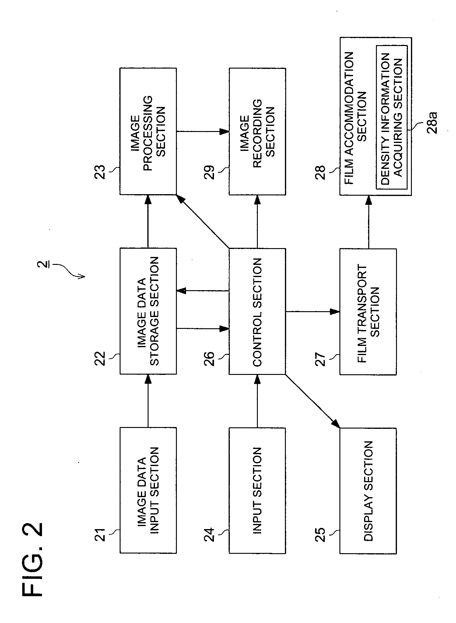
[0060]FIG. 1 is a conceptual diagram representing the system configuration of a medical image system 100 in the present embodiment. As shown in FIG. 1, the medical image system 100 is linked with medical image generation apparatuses 1a through 1e, medical image recording apparatuses 2a and 2b, HIS / RIS3, JOB manger 4, WS (Work Station) 5 and others through the network so that data can be transferred among them. Further, medical image generation apparatuses 1d and 1e, JOB manager 4 and WS5 constitute the CR network 10.

[0061] LAN (Local Area Network), WAN (Wide Area Network), the Internet and various forms of lines can be applied as the network N. Radio communications and infrared comm...


PUM


Abstract

A medical image system including: an image data input section for inputting image data including medical image data of a radiographed object and supplementary information; a plurality of outputting sections for outputting a medical image on the basis of the medical image data; a setting section for setting a setting density range or a setting brightness range of the medical image to be outputted on the basis of the inputted supplementary information; an information obtaining section for obtaining density information including a output density range or brightness information including a output brightness range which each of the outputting sections can output; a selecting section for selecting at least one of the outputting sections which can output the medical image data; and a transmitting section for transmitting the medical image data with the supplementary information to the selected outputting sections.

Description

FIELD OF THE INVENTION [0001] The present invention relates to a medical image system for outputting a medical image by converting the image signal inputted from a plurality of types of image signal sources into the image signal corresponding to the image signal source. BACKGROUND OF THE INVENTION [0002] In the prior art, various methods have come to be devised to capture medical radiographic image information, without using a radiographic film consisting of silver halide photosensitive material. For example, there has been widespread use of Computed Radiography apparatus (hereinafter abbreviated as “CR”) that utilizes the imaging plate mainly consisting of a stimulable phosphor to pick up photostimulated luminescence using excitation light after storing the radiographic image once, and applies photoelectric conversion to this light, thereby acquiring image signals. In recent years, a proposal has been made of a Flat Panel Detector (hereinafter abbreviated as “FPD”) for reading radi...

Claims


Application Information

Patent Timeline
IPC IPC(8): G06F19/00G06T5/40H04N1/32
CPCG06F19/3418H04N1/2361H04N1/32502H04N1/32512H04N2201/33307H04N1/33376H04N2201/0079H04N2201/0082H04N2201/3242H04N1/32529G16H30/40G16H30/20
Inventor KOMIYA, YUJIUMEKI, MAMORUNAKAZAWA, MASAYUKITODA, HARUYUKI
Owner KONICA MINOLTA MEDICAL & GRAPHICS INC
Why Eureka
Try Eureka