
Intratumoral delivery of dendritic cells

a dendritic cell and intravenous technology, applied in the direction of vertebrate antigen ingredients, biocide, antibody medical ingredients, etc., can solve the problems of inability to treat brain tumors in a single treatment, inconvenient treatment, and a large number of brain tumors, so as to reduce the severity of the complications, prevent the tumor from manifesting, and reduce the mass and/or size

Inactive Publication Date: 2004-03-25
CEDARS SINAI MEDICAL CENT
View PDF8 Cites 30 Cited by
  • Summary
  • Abstract
  • Description
  • Claims
  • Application Information

AI Technical Summary

Benefits of technology

[0012] The present invention provides a method and dendritic cell-based composition for treating a tumor. The method may include administering dendritic cells directly into the tumor itself or into the tissue surrounding or located nearby the tumor, and may be effective in the treatment of a tumor disposed in any location throughout the body of a mammal. The method and composition do not require the dendritic cells used therein to be primed ex vivo prior to inclusion in the composition or administration to a patient. The methods of the present invention may induce immune cell infiltration into a tumor.
[0034] In a preferred embodiment of the present invention, a mammal is treated with dendritic cells following or in conjunction with radiotherapy. While not wishing to be bound by any theory, it is believed that prior or simultaneous treatment with radiotherapy renders a dendritic cell vaccination more effective as is allows the dendritic cells to better process dying tumor cells. Similarly, a chemotherapy regimen administered either prior to or simultaneous with dendritic cell vaccination therapy that induces tumor cells to undergo apoptosis (i.e., programmed cell death) may be beneficial.
[0035] As used herein, "treating" a tumor includes, but is not limited to, ameliorating the tumor, lessening the severity of its complications, causing it to decrease in mass and / or size, preventing it from manifesting, preventing it from recurring, merely preventing it from worsening, or a therapeutic effort to effect any of the aforementioned, even if such therapeutic effort is ultimately unsuccessful.

Problems solved by technology

One particularly difficult group of tumors to treat are those that reside in and near the brain.
Treatment of brain tumors presents a number of problems, not the least of which being the dangers inherent in any surgical procedure involving regions of the brain and the tissue located nearby.
There is little room for error and the consequences of even a minor surgical mishap can be devastating to a patient; brain damage, or even death may result.
However, even commonly referenced medical authority suggests that patients with brain tumors be referred to centers specializing in investigative therapies; an indication that conventional modes of treatment are not overwhelmingly successful.
However, this treatment generally fails in substantially changing the outcome for a patient; median survival remains less than one year even with medical intervention.
Although this form of immunotherapy was well-tolerated by patients, therapeutic attempts implementing the same have thus far not led to the identification of a superior treatment or cure.
However, these studies further note that tumor cells are poor APCs; they do not efficiently internalize and process / present tumor antigens to T-cells. S. Constant et al., "Peptide and protein antigens require distinct antigen-presenting cell subsets for the priming of CD4+ T cells," J. Immunol. 154:4915-4923 (1995); D. Levin et al., "Role of dendritic cells in the priming of CD4+ lymphocytes to peptide antigen in vivo," J. Immunol. 151:6742-6748 (1993).
However, intracranial vaccination with cytokine-expressing cells is not clinically implemented, as intracranial cytokine expression presents a host of potential complications, such as the undesirable induction of an inflammatory response within the brain that is not targeted against tumor cells.
The key limitation in each of these dendritic cell-based vaccination strategies is their reliance on the acquisition of tumor tissue as a protein source for use in priming dendritic cells ex vivo.
However, priming the dendritic cells in this fashion precludes the use of this therapeutic modality in cases where tumor tissue cannot be readily obtained; a shortcoming frequently encountered with various types of tumors, since a variety of circumstances can render ex vivo priming either impractical or impossible.
For instance, a tumor may be surgically inaccessible, or the surgical manipulation thereof may present unreasonable danger to the health and safety of a patient.

Method used


Image

  • Intratumoral delivery of dendritic cells
  • Intratumoral delivery of dendritic cells
  • Intratumoral delivery of dendritic cells

Examples


example 1

Intracranial Dendritic Cell Vaccination of Brain Tumors

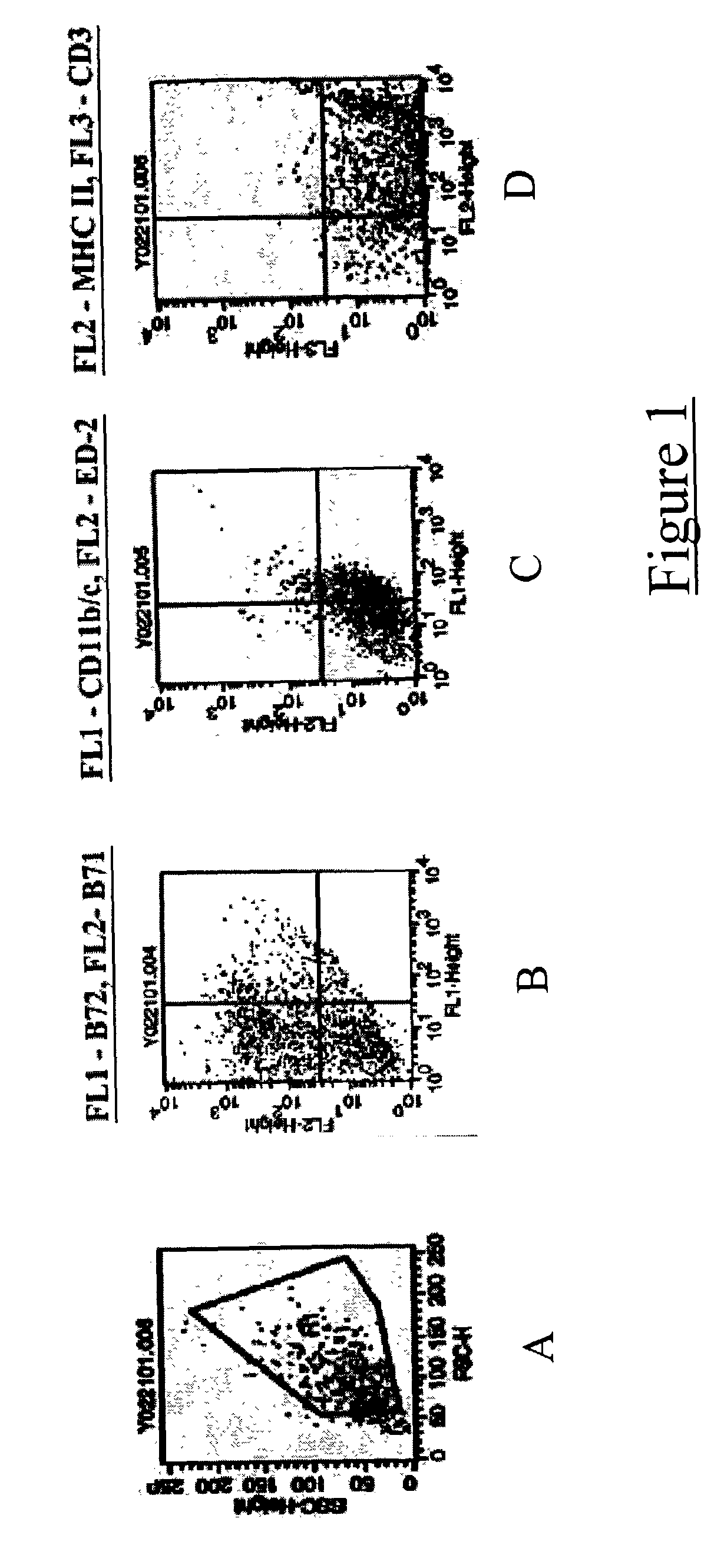
[0037] Bone marrow was harvested from the femurs and tibias of adult Fisher rats. Cells were plated in 24 well plates at a density of 1 million cells per well in RMPI 1640 medium (obtained from Gibco BRL; Gaithersburg, Md.; hereinafter "Gibco") in media containing GM-CSF and IL-4 (both available from R and D Systems; Minneapolis, Minn.; hereinafter "R and D"). Media was partially replenished every three days. After eight days, clusters of enlarged floating / partially adherent dendritic cells were apparent. These cells were collected separately and their phenotypic profiles assessed using flow immunocytometry. They were positive for MHC class II and B7 co-stimulatory molecules; thereby confirming that the cells were dendritic in nature (FIG. 1).

example 2

Dendritic Cells Inhibit Tumor Growth when Inoculated Intratumorally

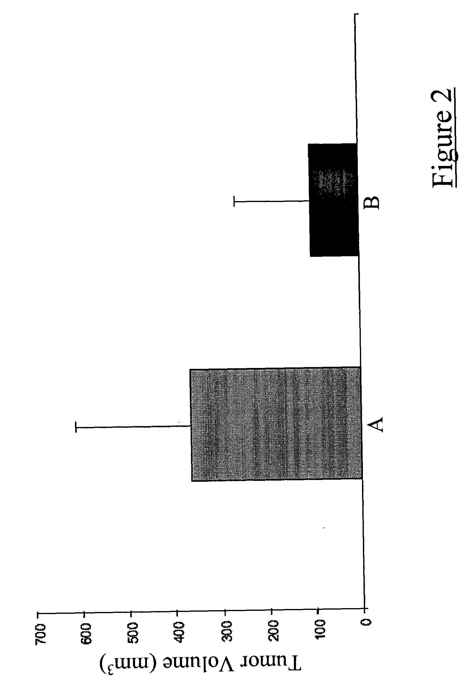
[0038] Dendritic cells were inoculated subcutaneously along with a mixture of irradiated and viable 9L glioma cells into the dorsum of the right foot of adult Fisher rats. Two weeks following this procedure, a second dose of dendritic cells was inoculated into each growing tumor. Eight weeks following the second dendritic cell vaccination, tumor sizes were measured using a precision caliper. Tumors were markedly smaller in animals that had received intratumoral dendritic cell vaccinations as compared to the control animals that received only saline inoculations (FIG. 2).

example 3

Dendritic Cell Vaccination Induces Immune Cell (T-cell) Infiltration into Brain Tumors

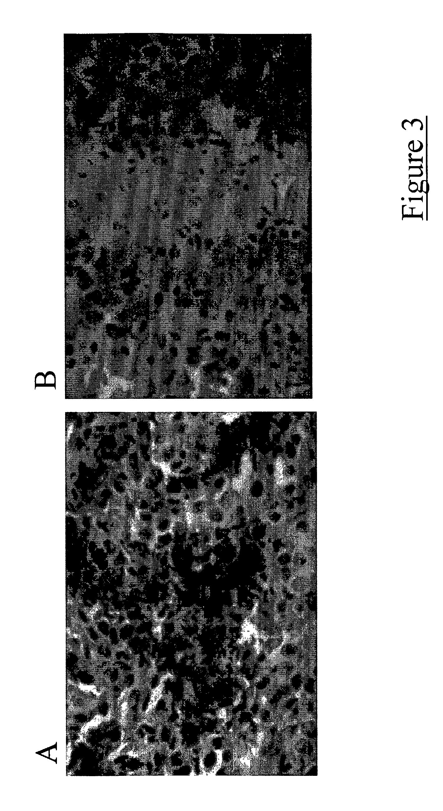
[0039] Dendritic cells were inoculated intracranially along with a mixture of irradiated and viable 9L glioma cells into the right corpus striatum (basal ganglia) of adult Fisher rats. Two weeks following this procedure, a second dose of dendritic cells was inoculated into each growing tumor. Two weeks following the second dendritic cell vaccination, animals were euthanized and their brains harvested. The brains were immediately frozen and sectioned on a cryostat (available from Janis Research Company, Inc.; Wilmington, Mass.). Slide mounted sections were stained for T-cell markers (i.e., CD4 and CD8). Tumors from dendritic cell vaccinated animals displayed increased quantities of infiltrating T-cells as compared to tumors from control animals that received only saline inoculations (FIG. 3).


PUM

PropertyMeasurementUnit
Timeaaaaaaaaaa
Volumeaaaaaaaaaa

Abstract

Methods included herein describe the treatment of a tumor by administering dendritic cells either directly into the same or into its surrounding tissue. Further methods describe the induction of immune cell infiltration into tumors and the treatment of tumors with unprimed dendritic cells by administering dendritic cells in a similar fashion. The methods of the present invention are particularly advantageous in the treatment of brain tumors and other solid tumors disposed throughout the body of a mammal that are difficult or impossible to treat by conventional surgical means. Dendritic cell-based compositions effective in the treatment of such tumors are also described.

Description

[0001] The present invention relates to methods of treating a tumor by administering dendritic cells and compositions effective for the same. More specifically, the method involves administering dendritic cells directly into a tumor or its surrounding tissue, the tumor being located in the body of a mammal. The compositions are dendritic cell-based.[0002] Cancer remains one of the leading causes of death in the United States and around the world. Various forms of cancer are differentially treated, depending in part on the location of a tumor targeted for treatment. One particularly difficult group of tumors to treat are those that reside in and near the brain. Treatment of brain tumors presents a number of problems, not the least of which being the dangers inherent in any surgical procedure involving regions of the brain and the tissue located nearby. There is little room for error and the consequences of even a minor surgical mishap can be devastating to a patient; brain damage, or...

Claims


Application Information

Patent Timeline
IPC IPC(8): A61K39/00
CPCA61K39/0011A61K2039/5154A61K2039/5152A61K39/4644A61K39/4615A61K39/4611A61K39/4622A61K35/15
Inventor YU, JOHNBLACK, KEITHEHTESHAM, MONEEB
Owner CEDARS SINAI MEDICAL CENT
Learn More