
Efficient methods for assessing and validating ecandidate protein-based therapeutic molecules encoded by nucleic acid sequences of interest

Inactive Publication Date: 2003-07-03
MEDGENICS INC +1
View PDF12 Cites 17 Cited by
  • Summary
  • Abstract
  • Description
  • Claims
  • Application Information

AI Technical Summary

Problems solved by technology

From this point of view, it is just as important to invalidate a protein-based drug, which does not show sufficient physiological / therapeutic effect.
However, in vitro study can give only limited information, and animal-based systems must be used to reach operative conclusions regarding the biological / physiological effect / activity of the protein or nucleic acid sequence.
However, current in vivo approaches require lengthy and expensive procedures for protein production, purification and formulation, all before administration to an animal is even possible.
It is often difficult, time consuming, costly, and sometimes even impossible to achieve high-level expression of a given recombinant protein.
Each of the above-described hosts has limitations in terms of either the amount of protein expressed, or other aspects of the protein, which relate to its activity in the intended use.
For example, proteins expressed in bacterial cells, which are the easiest to manipulate, are often maintained in a non-secreted manner inside the bacterial cell and more specifically are localized within inclusion bodies from which it is oftentimes difficult to isolate and purify them.
Furthermore, a bacterial cell cannot provide to the protein many of the post-translational modifications (such as glycosylation and the accurate folding of the protein) that may be required for its biological activity.
On the other hand, eukaryotic protein production systems may result in inaccurate post-translational modification.
In certain circumstances, an expressed recombinant protein might be toxic to the host cells, which further prevents production of reasonable amounts for assessing that protein.
Development of a purification scheme is a very lengthy process.
Often, it is necessary to sustain substantial production losses with very low yields in order to obtain recombinant protein of the necessary purity.
The process of developing an appropriate formulation is time consuming, difficult, and costly, as well.
Furthermore, even formulated, purified recombinant proteins have a finite shelf life due to maintenance and storage limitations; often requiring repeated purification and formulation of more protein.
Batch-to-batch variation encountered in such an approach may complicate the data obtained in animal studies using these proteins.
All the above-described protein production techniques are very lengthy and costly, and frequently do not yield sufficient, biologically active amounts of the desired protein to enable the intended required analysis in vitro and in vivo.
However, several as yet insurmountable limitations plague their efficient application.
Additionally, because of the requirement for retroviral integration within the subject's genome, the vector can only be used to transduce actively dividing tissues.
Further, many retroviruses have limited host tissue specificity and cannot be employed to transduce more than a few specific tissues of the subject.
Other DNA based viral vectors suffer limitations as well, in terms of their inability to sustain long-term transgene expression; secondary to host immune responses that eliminate virally transduced cells in immune-competent animals (Gilgenkrantz et al., Hum.
These combined limitations result in inconsistent recombinant gene product expression, and a difficulty in determining accurate expression levels of the recombinant product, and little opportunity for prolonged in vivo expression.
Centrifugation did not enhance, and may even have hindered efficient transgene incorporation.
Deleterious physiological effects may involve, but are not limited to, destructive invasion of tissues, growth at the expense of normal tissue function, irregular or suppressed biological activity, aggravation or suppression of an inflammatory or immunologic response, increased susceptibility to other pathogenic organisms or agents, and undesirable clinical symptoms such as pain, fever, nausea, fatigue, mood alterations, and other features.
Among the more difficult tasks in drug design is optimization of particular compounds once a therapeutic effect is discovered.
Random testing in whole animals is a costly, time consuming procedure as outlined hereinabove.
These methods, however, do not address functional relationships between multiple proteins simultaneously, in vivo, in whole animal systems.
Applications of in vivo introduction of genetic sequences for in vivo production of recombinant gene products, (and in cases where the construct provides for the production of a product that is otherwise defective or absent the methodology is otherwise referred to as "gene therapy"), have significant limitations.
Because of the requirement for integration into the subject genome, the retrovirus vector can only be used to transduce actively dividing tissues, posing another limitation to the method application.
Further, many retroviruses have limited host tissue specificity and cannot be employed to transduce more than a few specific tissues of the subject (Kurian K M, Watson C J, Wyllie A H.
Adenoviral vectors have been another preferred vector of choice for gene therapy attempts, but they too are limited in potential therapeutic use for several reasons.
First, due to the size of the El deletion and to physical virus packaging constraints, first generation adenovirus vectors are limited to carrying approximately 8.0 kb of transgene genetic material.
While this compares favorably with other viral vector systems, it limits the usefulness of the vector where a larger transgene is required.
Second, infection of the E1-deleted first generation vector into packaging cell lines leads to the generation of some replication competent adenovirus particles, because only a single recombination event between the E1 sequences resident in the packaging cell line and the adenovirus vector genome can generate a wild-type virus.
Therefore, first-generation adenovirus vectors pose a significant threat of contamination of the adenovirus vector stocks with significant quantities of replication competent wild-type virus particles, which may result in toxic side effects if administered to a gene therapy subject (Rubanyi, G. M. (2001) Mol Aspects Med 22(3): 113-42.
The most difficult problem with most vectors employed, including adenovirus vectors is their inability to sustain long-term transgene expression, secondary to host immune responses that eliminate virally transduced cells in immune-competent animals (Gilgenkrantz et al., (1995) Hum.

Method used


Image

  • Efficient methods for assessing and validating ecandidate protein-based therapeutic molecules encoded by nucleic acid sequences of interest
  • Efficient methods for assessing and validating ecandidate protein-based therapeutic molecules encoded by nucleic acid sequences of interest
  • Efficient methods for assessing and validating ecandidate protein-based therapeutic molecules encoded by nucleic acid sequences of interest

Examples


example 1

[0230] In Vitro Micro-Organ Expression of Murine Erythropoietin

[0231] Material and Experimental Methods

[0232] Preparation of Human Skin Micro-Organs

[0233] Approval for experiments utilizing human skin was obtained from the Rambam Hospital, Israel, according to standards approved by the Helsinki committee. A section of 1.4-1.5 mm human female skin thickness (depth) was aseptically removed from the abdomen according to standard operating procedures. The biopsy tissue was treated with a hypochloride solution (10% Milton solution), for 7 minutes followed by 3 washes with 20 ml DMEM for 10 minutes each. Following treatment, the tissue was further sectioned with a tissue chopper (TC-2 chopper, Sorval, Du-pont instruments). Tissue sectioning into 300 .mu.m width explants was conducted under sterile conditions. The resulting micro-organs (MOs) were placed individually within wells of a 48-well micro-plate containing 400 .mu.l per well of DMEM (Biological Industries --Beit Haemek) in the abs...

example 2

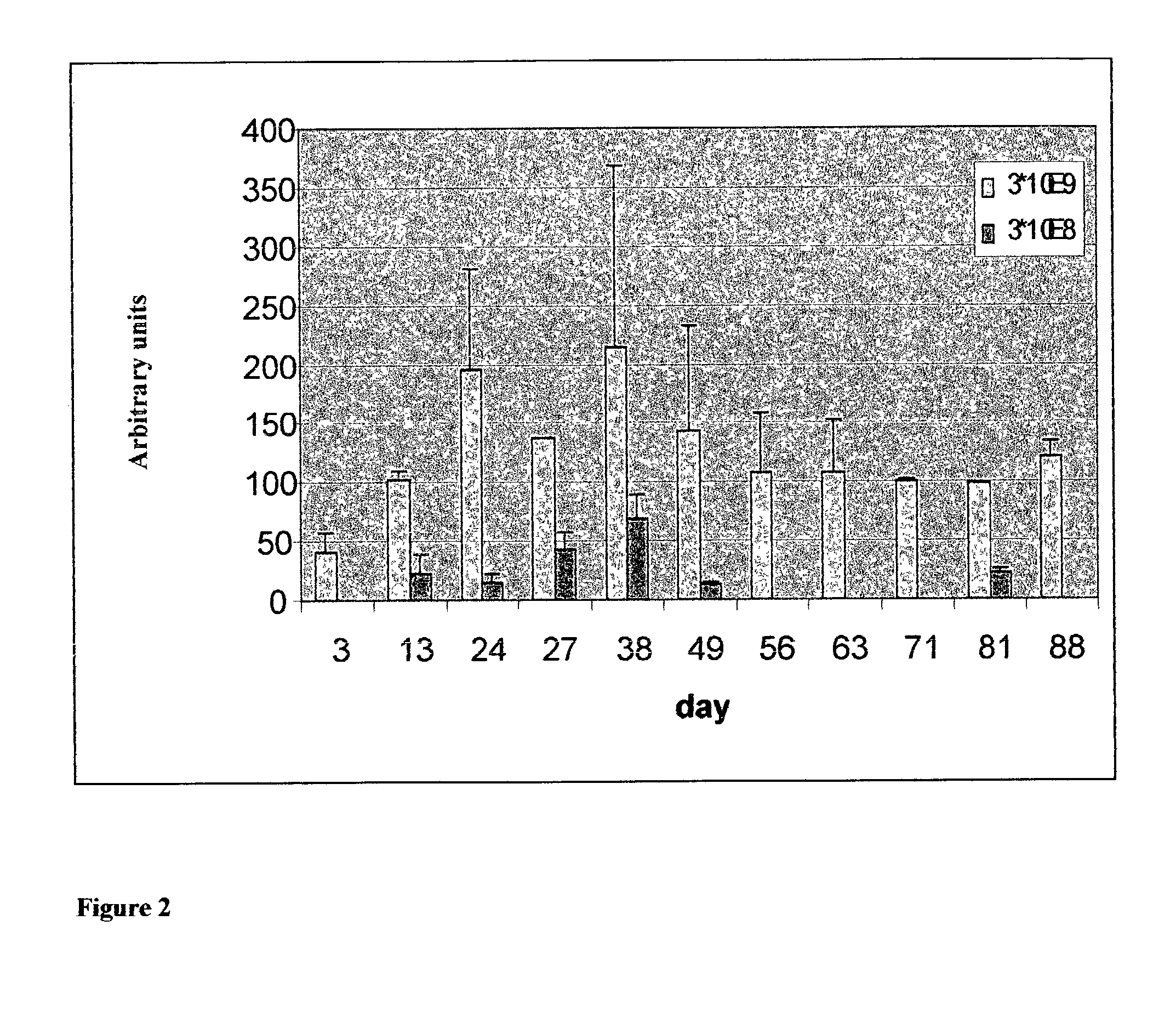
[0251] In vivo Micro-Organ Expression of Murine Interferon-.alpha.

[0252] Material and Experimental Methods

[0253] Construct Preparation:

[0254] The commercially available vector comprising strain 5 of the adenovirus expressing murine interferon a off the cytomegaloviral promoter (designated Ad5-CMV / mIFN.alpha.) and a vector comprising strain 5 of the adenovirus expressing the .beta.-galactosidase gene, (designated Ad5-CMV / LacZ), used as a control, were both purchased from Q-Biogene (Carisbad, Calif., USA).

[0255] Ad5-CMV / mIFN .alpha. micro-organ implantation:

[0256] Male and female SCID mice weighing around 25 grams were anaesthetized with 140 ul of diluted Ketast (ketamine HCl) (400 .mu.l Ketast and 600 .mu.l saline) and Ad5-CMV / mIFN .alpha. expressing MOs were implanted subcutaneously, 14 days following MO transduction.

[0257] Assessment of in vitro Protein Production:

[0258] Media was removed from each well every 2-3 days and assayed via ELISA for the presence of secreted mIFN.alpha. (...

example 3

[0271] Implanted Microorgans Maintain Structural Integrity Over Time

[0272] Material and Experimental Methods

[0273] Preparation of Murine Lung Micro-Organs:

[0274] Entire lungs were removed from several C57B1 / 6 mice and then lower right or left lobes of the lungs were aseptically dissected. The tissue was further sectioned with a tissue chopper (TC-2 Tissue sectioning, Sorval Du-pont instruments) into 300 .mu.m width explants, under sterile conditions. The resulting micro-organs (MOs) were placed within wells of a 48-well micro-plate containing 400 .mu.l of DMEM (Biological Industries--Beit Haemek) in the absence of serum, per well, and incubated under a 5% CO2 atmosphere, at 37.degree. C. for 24 hours. Wells were visualized under a binocular (Nikon-SMZ 800) microscope and micro-organs were photographed, accordingly.

[0275] Experimental Results

[0276] MOs Maintain Macroscopic Integrity during Long-Term Sub-Cutaneous Implantation

[0277] Mouse lung MO's were prepared similarly to human ski...


PUM

PropertyMeasurementUnit
Lengthaaaaaaaaaa
Lengthaaaaaaaaaa
Timeaaaaaaaaaa

Abstract

A method of determining at least one quantitative or qualitative pharmacological, physiological and / or therapeutic, parameter or effect of a recombinant gene product in vivo, the method comprises (a) obtaining at least one micro-organ explant from a donor subject, the micro-organ explant comprising a population of cells, the micro-organ explant maintaining a microarchitecture of an organ from which it is derived and at the same time having dimensions selected so as to allow diffusion of adequate nutrients and gases to cells in the micro-organ explant and diffusion of cellular waste out of the micro-organ explant so as to minimize cellular toxicity and concomitant death due to insufficient nutrition and accumulation of the waste in the micro-organ explant, at least some cells of the population of cells of the micro-organ explant expressing and secreting at least one recombinant gene product; (b) implanting the at least one micro-organ explant in a recipient subject; and (c) determining the at least one quantitative or qualitative pharmacological, physiological and / or therapeutic, parameter or effect of the recombinant gene product in the recipient subject.

Description

[0001] This application is a continuation of PCT / IL02 / XXXXX, filed Jul. 7, 2002, having the same title and identified by Attorney Docket No. 02 / 23844, which claims the benefit of priority from U.S. Provisional Patent Application No. 60 / 303,337, filed Jul. 9, 2001.FIELD AND BACKGROUND OF THE INVENTION[0002] The present invention relates to methods of rapid assessment and validation of candidate protein-based therapeutic molecules encoded by nucleic acid sequences of interest. The present invention also relates to methods of determining at least one quantitative or qualitative pharmacological, physiological and / or therapeutic parameter or effect of an expressed recombinant gene product in vitro or in vivo. More particularly, the present invention relates to a method of determining these effects in an in vivo system utilizing micro-organs as a means of expressing nucleic acids of interest.[0003] The human genome project has provided the scientific world and the biotechnological and pha...

Claims


Application Information

Patent Timeline
IPC IPC(8): C12N15/10
CPCC12N15/1034C12N15/1079C12N15/1055
Inventor GARFINKEL, LEONARD I.PEARLMAN, ANDREW L.MITRANI, EDUARDO N.
Owner MEDGENICS INC
Learn More