Eureka AIR delivers breakthrough ideas for toughest innovation challenges, trusted by R&D personnel around the world.

Vacuum-assisted drug delivery device and method

a technology of vacuum injection and drug delivery, which is applied in the direction of medical devices, infusion needles, medical applicators, etc., can solve the problems of eye infections, eye infections, and eye infections that are difficult to treat with traditional methods

Active Publication Date: 2022-06-07
SEROS MEDICAL
View PDF19 Cites 1 Cited by
  • Summary
  • Abstract
  • Description
  • Claims
  • Application Information

AI Technical Summary

Benefits of technology

[0021]In an aspect of this invention, the drug delivery device further comprises a proximal or backward flow inhibitor, such as a valve or stepped diffusor, to prevent or reduce the fluid in the drug reservoir from being withdrawn from the drug reservoir when a vacuum is being created by the vacuum source.
[0039]In an aspect of this invention, upon delivery of an effective amount of riboflavin to ocular tissue, the eye may be exposed to radiation such as ultraviolet light to crosslink tissue exposed to riboflavin, the riboflavin serving as a photosensitizer to create new covalent crosslinks between collagen strands to increase corneal stiffness.
[0047]An aspect of this invention is a method for delivering a drug non-invasively through the epidermis, comprising: placing a surface of a drug reservoir in contact with a target area of the epidermis wherein the surface of the drug reservoir is configured to form an air-tight contact between the drug reservoir surface and the epidermal surface and wherein a fluid in the drug reservoir is in fluid communication with the epidermal surface; and creating a vacuum within the drug reservoir to accelerate diffusion of the fluid in the drug reservoir through the epidermis and into target tissue beneath the epidermis.
[0052]In an aspect of this invention, the vacuum is pulsed to further improve diffusion of the fluid through the epidermis.
[0058]An aspect of this invention is a method for delivering a fluid noninvasively through a surface membrane of an internal organ, comprising: accessing the internal organ; placing a surface of a drug reservoir in contact with a target area on the surface membrane of the internal organ wherein the surface of the drug reservoir is configured to form an air-tight contact between the drug reservoir surface and the internal organ membrane surface and wherein a fluid in the drug reservoir is in fluid communication with the internal organ surface; and creating a vacuum within the drug reservoir to accelerate diffusion of the fluid in the drug reservoir through the surface of the internal organ and into target tissue beneath.

Problems solved by technology

Delivery of drugs that provide effective therapeutic concentrations to targeted tissues, with minimal side effects, is a major challenge because of physiological and anatomical barriers.
As a result of this natural protection, the eye can be extremely difficult to treat.
This technique poses some risk to the patient, especially when it is necessary to perform multiple injections, such as injecting the drug Avastin into the vitreous for treatment of age-related macular degeneration.
Another type of ophthalmic drug delivery, which is painful to the patient and can sometimes lead to eye infections, is the delivery of drug directly into the cornea.
Moreover, the recovery period is painful to the patient, and there is also risk of haze and scar formation.
KCS, also known as dry eye, is an eye condition caused by inflammation of the cornea and conjunctiva resulting from lack of tear production, causing dryness and pain in those tissues.
The KCS condition causes ocular irritation, loss of vision, ocular surface diseases, and the risk of corneal ulceration.
Oral ingestion and injection of drugs, with their respective systemic side effects, are not always safe and practical.
For example, a significant obstacle to dermal and transdermal drug delivery is the resilient barrier that the epidermal layers of the skin, primarily the stratum corneum, presents for the diffusion of exogenous chemical agents.
Another challenge is the delivery of various therapeutic agents deeper within the skin, such as into the sudoriferous and sebaceous glands or at the base of hair follicles.
However, very few drugs are suitable for topical delivery, with passive penetration through skin normally being limited to small molecules (<500 Da), which are neutral and relatively lipophilic.

Method used


Image

  • Vacuum-assisted drug delivery device and method
  • Vacuum-assisted drug delivery device and method
  • Vacuum-assisted drug delivery device and method

Examples


Embodiment Construction

Brief Description of the Drawings

[0068]The figures herein are presented to aid in the understanding of the invention and are not intended nor should they be construed as limiting the scope of this invention in any manner. Thus, while the figures as such are directed to a device for vacuum-assisted non-intrusive delivery of a drug to the eye and the skin, it is understood that the figures may be modified in ways based on the disclosures herein to adapt to the vacuum-assisted, non-invasive delivery of drugs through the outer layer of any organ.

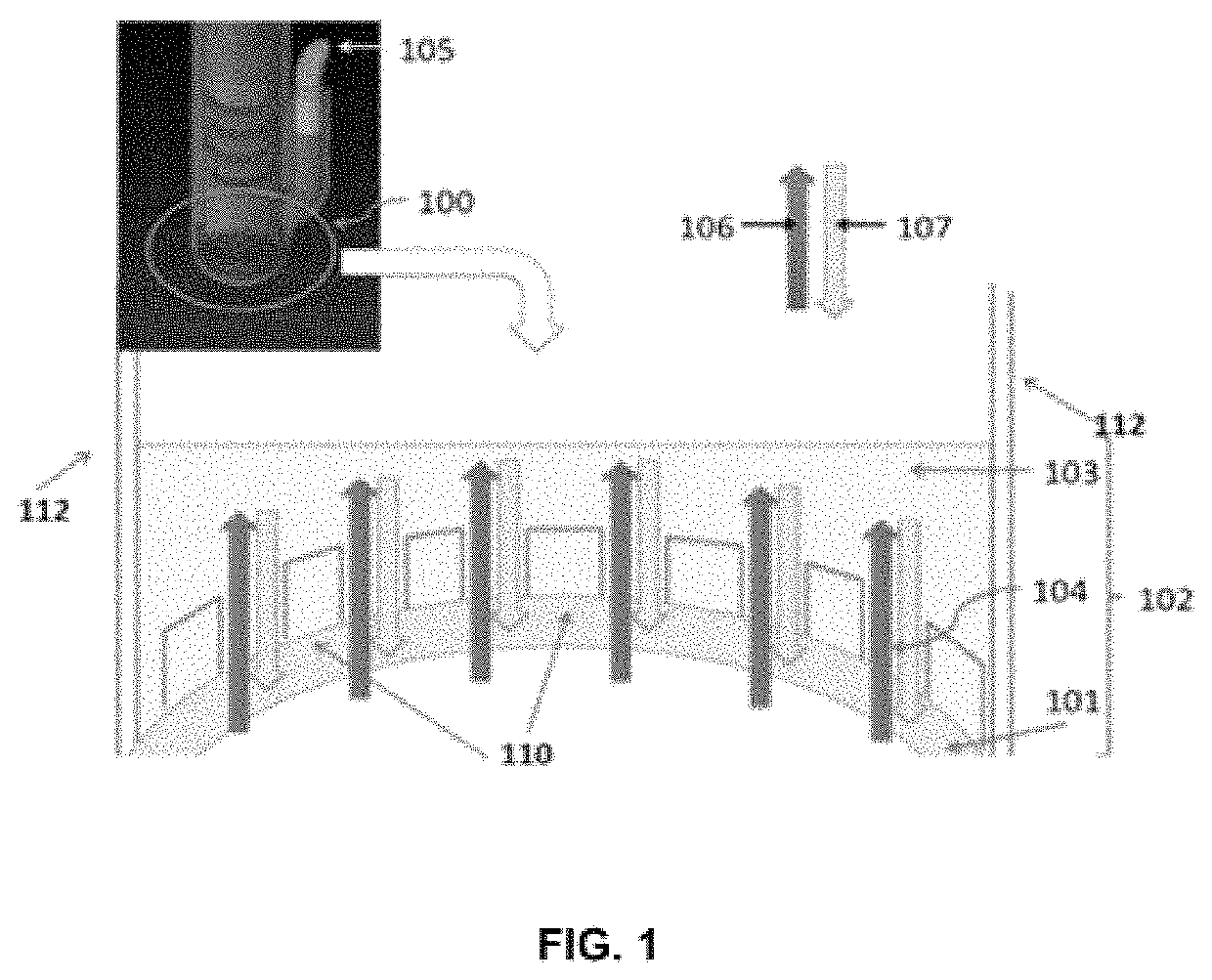
[0069]FIG. 1 is a schematic setting forth the mechanism of action of a vacuum delivery system.

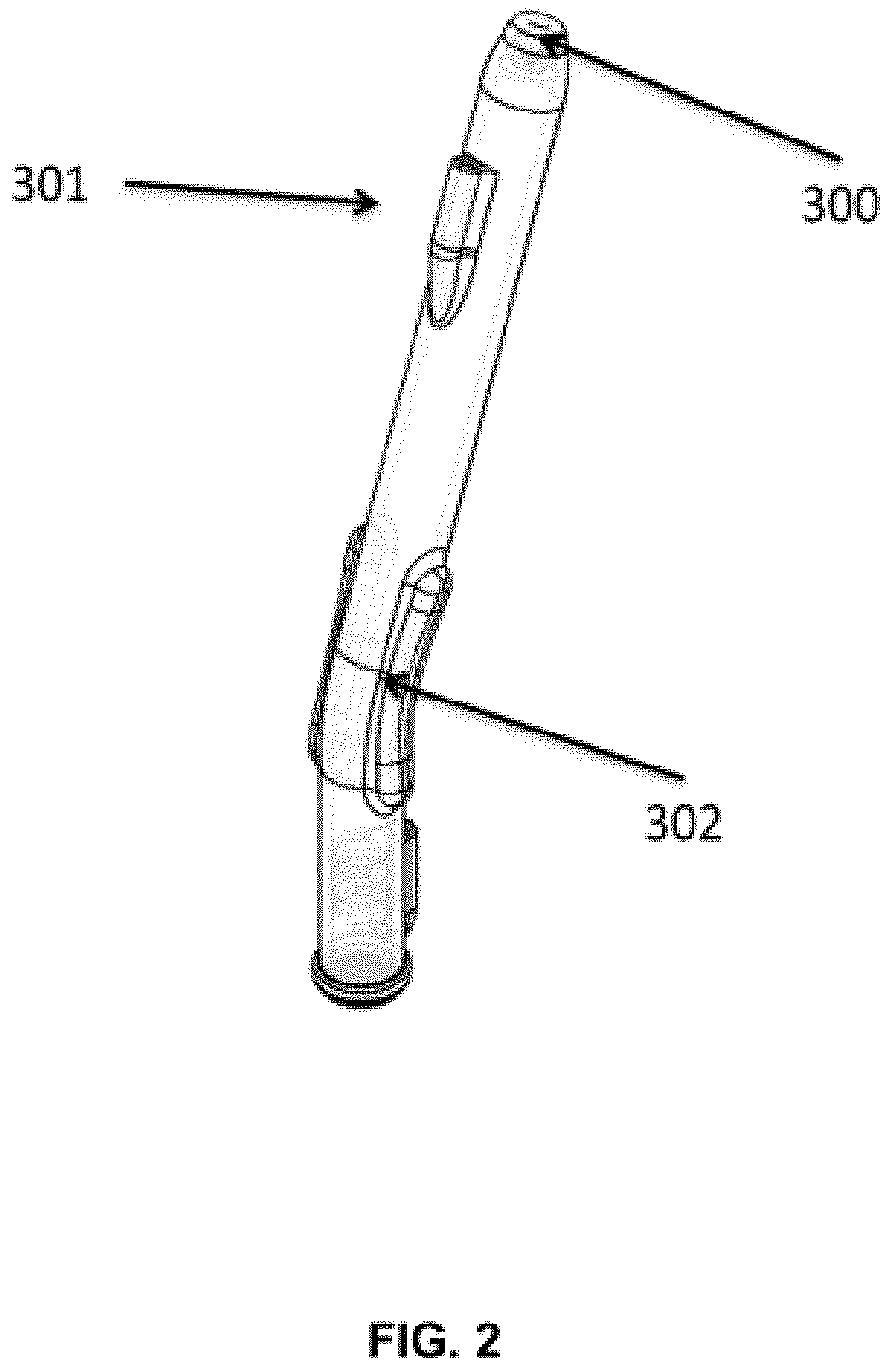
[0070]FIG. 2 is a drawing of the side of the device. Handles are on the radii of the curve. The vacuum tube is on the proximal end of the handle.

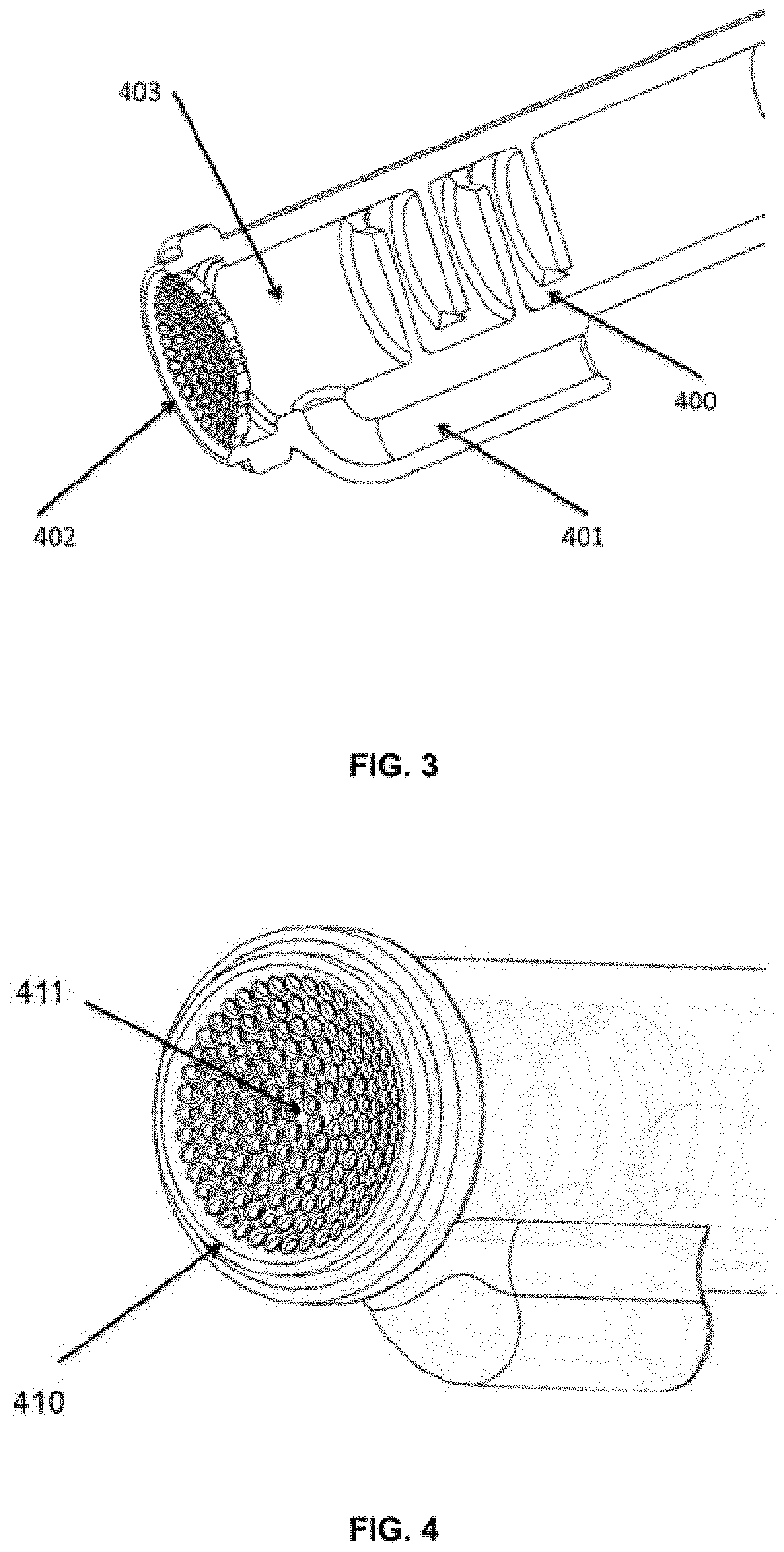
[0071]FIG. 3 is a cut-out view through the center axis. The proximal or backward flow inhibitor or stepped diffusor is shown which keeps solutes from being retracted by the applied vacuum. The solute inlet is j...


PUM


Abstract

This invention relates to a device and method for the vacuum-assisted delivery of drugs through an intact surface membrane of an organ.

Description

[0001]This application claims the benefit of U.S. Application No. 62 / 244,678 filed Oct. 21, 2015, which is incorporated by reference herein.[0002]This application is also a continuation-in-part of application Ser. No. 14 / 694,703 filed Apr. 23, 2015 which claims the benefit of U.S. Application No. 61 / 983,409 filed Apr. 23, 2014 and U.S. Application No. 62 / 138,330 filed Mar. 25, 2015, all of which are incorporated by reference herein.FIELD OF THE INVENTION[0003]This invention relates to a non-invasive drug delivery device and method. In particular, it relates to a device for vacuum-assisted drug delivery across unmodified tissue membranes, such as the skin and the epithelium of the eye.BACKGROUND OF THE INVENTION[0004]There are a multitude of drug delivery methods. Most current methods of drug delivery entail an initial delivery through oral ingestion, inhalation, transmucosal absorption, intravascular injection or intravenous modalities. Some drugs can now be delivered with new techn...

Claims


Application Information

Patent Timeline
Patent Type & Authority Patents(United States)
IPC IPC(8): A61F9/00A61M37/00A61M5/158A61M5/142A61M1/00A61M35/00
CPCA61M37/00A61F9/0008A61F9/0017A61M1/00A61M5/14248A61M5/158A61M35/003A61M2037/0007A61M2210/0612
Inventor MYUNG, DAVIDMANCHE, EDWARD E.EATON, DONALD J.ABBATE, ANTHONY J.
Owner SEROS MEDICAL
Why Eureka
Eureka Blog
Learn More