
Methods for improving proliferation and stemness of limbal stem cells

a stem cell and proliferation technology, applied in the field of stem cell proliferation and stem cell proliferation, can solve the problems of corneal epithelium renewal failure, serious visual impairment, and risk of lscd in the donor eye, and achieve the effect of improving the colony forming efficiency (cfe) of lscs

Active Publication Date: 2021-01-19
THE CATHOLIC UNIV OF KOREA IND ACADEMIC COOP FOUND
View PDF2 Cites 0 Cited by
  • Summary
  • Abstract
  • Description
  • Claims
  • Application Information

AI Technical Summary

Benefits of technology

The method significantly enhances the proliferation and stemness of limbal stem cells, improving their colony-forming efficiency and yield, which can lead to increased successful treatment rates for LSCD patients by providing a higher number of viable stem cells for transplantation.

Problems solved by technology

In LSC deficiency (LSCD), the migration of basal corneal epithelial cells onto a surface of the cornea and the migration of corneal epithelial cells on the periphery portions into the center thereof are reduced, and the loss of corneal epithelial cells is thus greater than the proliferation, resulting in a failure in renewal of the corneal epithelium.
When the cornea has opacity without maintaining its transparency because of external injuries, serious inflammations or congenital causes, even though all of the other functions of the eye as well as optic nerve are normal, a serious visual impairment is made.
In the case of autologous limbal tissue transplantation, a method for transplanting the same size of limbal tissue which has been taken from the other eye is used, which, however, has a risk of development of LSCD in the donor eye.
Meanwhile, in the case of the transplantation of homologous limbal tissues donated from a dead body, patients are suffering from side effects caused by the constant use of a systemic immunosuppressant, and the death of a significant amount of stem cells frequently occurs due to limbal graft rejection even when the systemic immunosuppressant is used.
As described above, while LSCD is constantly increasing, a proper treating method which is effective for patients with less complications has not been developed yet.
However, the isolation of the LSCs is very difficult because the number of the LSCs are less than 1% and, the LSCs are embedded in a basal membrane of the multilayer limbal epithelium.
To solve such a problem, conventionally, methods for culturing of multipotent stem cells isolated from the corneal limbus have been reported (Korean Patent No. 10-0931887), but there is no efficient method for clinical and practical uses.

Method used


Image

  • Methods for improving proliferation and stemness of limbal stem cells
  • Methods for improving proliferation and stemness of limbal stem cells
  • Methods for improving proliferation and stemness of limbal stem cells

Examples


example 1

on and Methods for Experiment

[0052]1-1. Preparation of Limbal Tissue

[0053]Post-keratoplasty discards of human corneal-limbal tissues from unidentifiable cadavers were obtained from an eye bank, and in the experiment, only corneal limbal tissue (1) isolated from a donor ranging in age from 30 to 65 years at death, (2) isolated within 12 hours after the death of the donor, (3) stored in Optisol (Bausch & Lomb, USA) less than 48 hours after the tissue harvest, and (4) isolated from a donor who had not been infected by human immunodeficiency virus (HIV), hepatitis B or C virus, Epstein-Barr virus, or syphilis was used.

[0054]The corneal limbal tissue satisfying the above-described conditions was splitted into 8 equal quarters using a surgical instrument on a horizontal sterile-air-flow hood, and the conjunctival edge of each quarter was immersed shortly in 1% trypan blue (Sigma, USA) to stain conjunctival stroma, and all remnants of conjunctiva were carefully removed using conjunctival s...

example 2

ve Analysis of Explant Outgrowth Proliferation Ability by Regulation of Wnt / β-Catenin Signaling in Limbal Explant Cultures

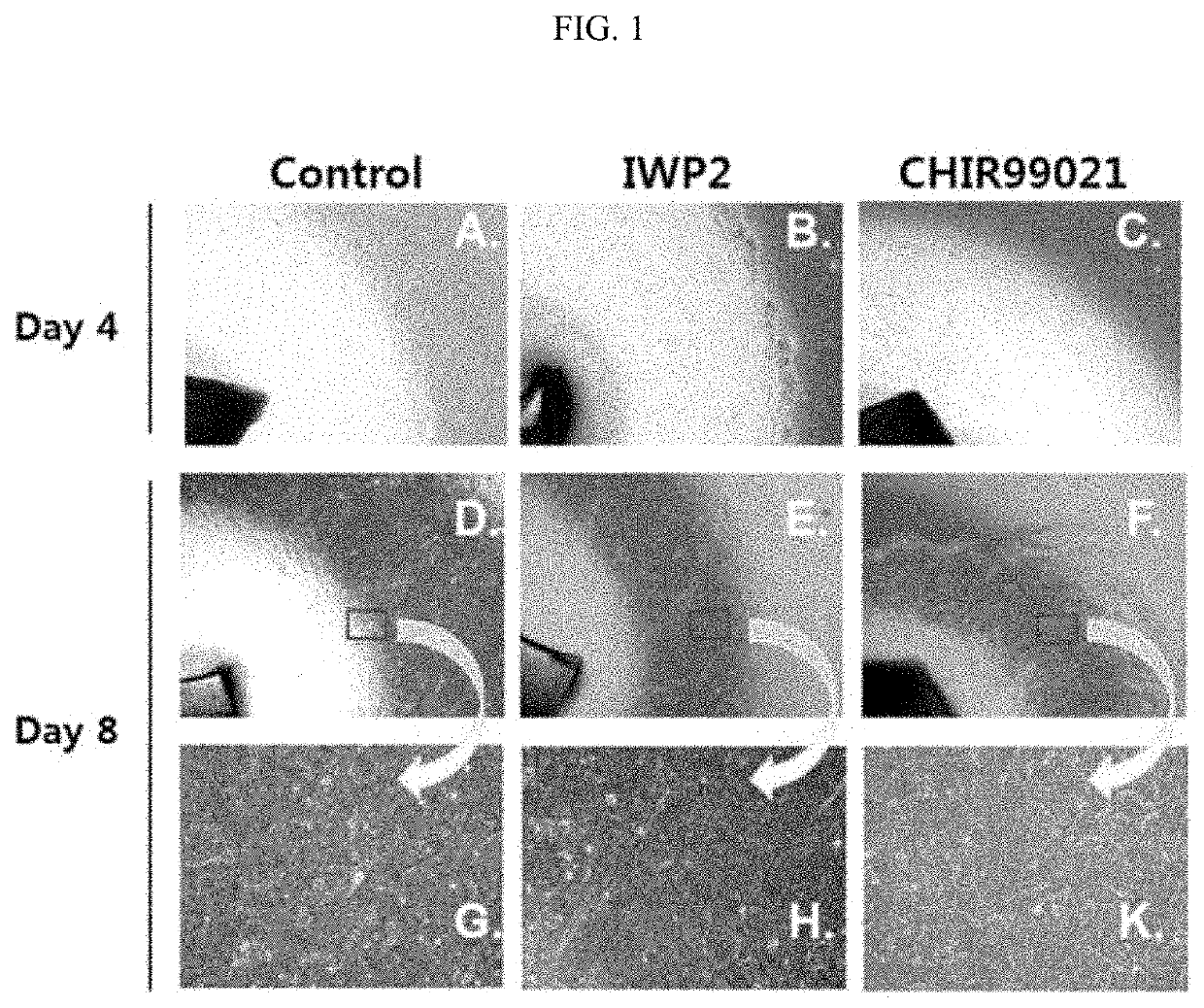
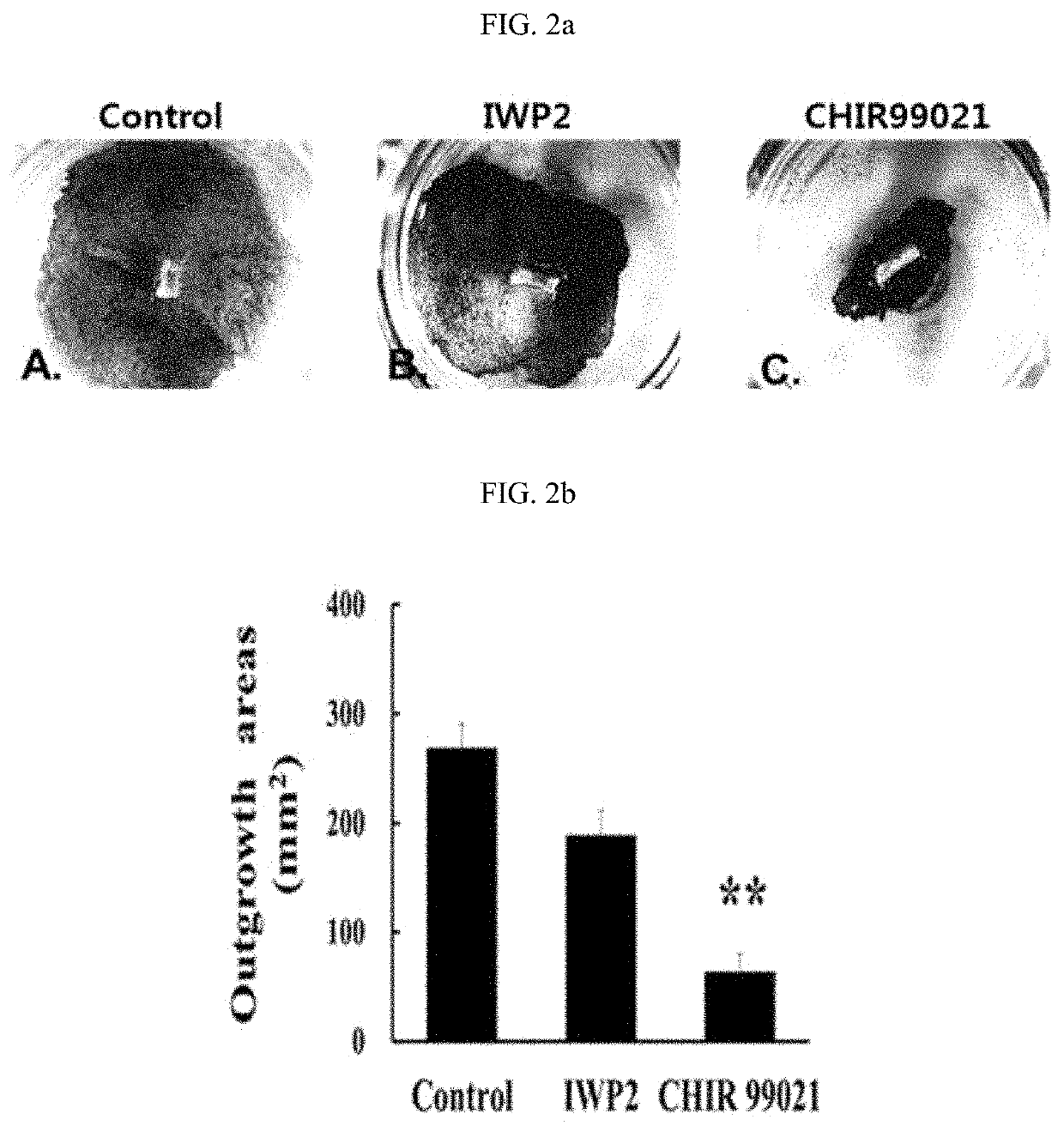
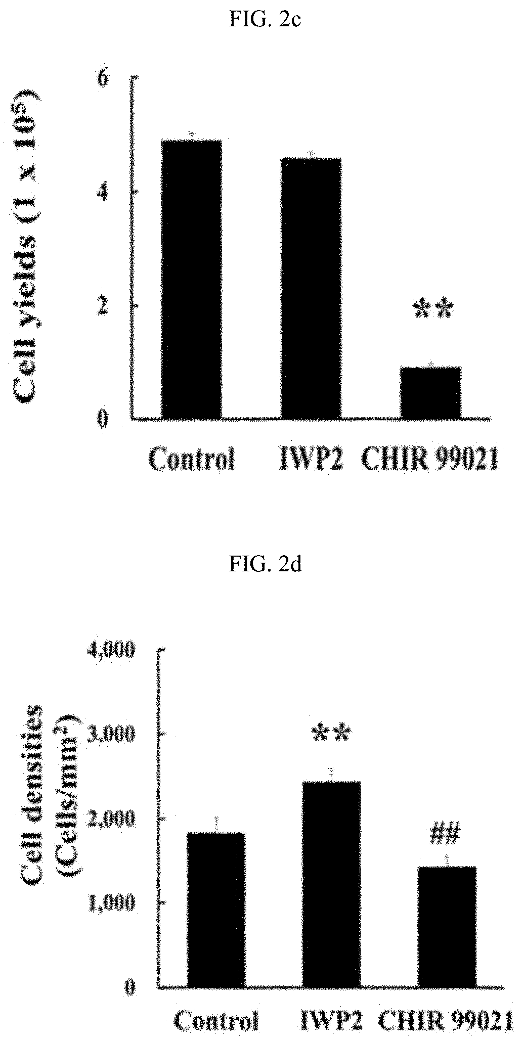
[0065]To identify effects of the Wnt / β-catenin signaling on the limbal explant outgrowth cultures, cells were cultured by adding 10 μM of the Wnt inhibitor IWP2 or 3 μM of the Wnt signaling agonist CHIR99021 as a GSK3 inhibitor to a medium under the culture conditions for a human limbal explant cultures according to the methods described in Examples 1-1 and 1-2, and explant outgrowth proliferation was observed using a phase contrast microscope on day 4 and 8.

[0066]The Wnt inhibitor IWP2 inhibited the Wnt signaling through inactivation by inhibiting an acetyltransferase activity of the porcupine protein involved in acylation of a Wnt protein, resulting in inhibition of Wnt / β-catenin signaling. Meanwhile, the GSK3 inhibitor CHIR99021 inhibited GSK3, which is involved in degradation of the β-catenin protein in cells, thereby increasing a level of the β-catenin in th...

example 3

ve Analysis of LSC Ratio by Regulation of Wnt / β-Catenin Signaling in Limbal Explant Cultures

[0071]In order to identify effects of the Wnt / β-catenin signaling on LSCs present in the explant during the limbal explant culture, cells were cultured by adding 10 μM of the Wnt inhibitor IWP2 or 3 μM of the GSK3 inhibitor CHIR99021 to a medium for a human limbal explant cultures according to the method described in Examples 1-1 and 1-2 and then subjected to flow cytometry according to the method described in Example 1-4, resulting in detection of the side population of JC1low cells in the explant cultures.

[0072]As a result, as shown in FIG. 3, it was identified that the explant cultured by IWP2 treatment showed the highest JC1 exclusion cell cohort (JC1low), which was 29.6±2.6%, and the CHIR99021-treated group showed the lowest JC1 exclusion cell cohort, which was 10.9±1.3%. Also, the explant cultured by IWP2 treatment showed the highest percentage in a lower left quadrant (LLQ) exhibiting ...


PUM

PropertyMeasurementUnit
thicknessaaaaaaaaaa
sizeaaaaaaaaaa
thicknessaaaaaaaaaa

Abstract

Provided are methods for improving the proliferation and stemness of limbal stem cells (LSCs) by adding a Wnt inhibitor to a medium for limbal explant cultures. When human limbal explants are cultured according to the methods, Wnt / β-catenin signaling is inhibited, resulting in improvement of LSC proliferation, and improvement in the stemness of LSCs, and thus the LSCs may be obtained in high yield.

Description

CROSS-REFERENCE TO RELATED APPLICATION[0001]This application claims priority to and the benefit of Korean Patent Application No. 10-2016-0037741, filed on Mar. 29, 2016, the disclosure of which is incorporated herein by reference in its entirety.[0002]The present invention was undertaken with the support of Analysis of the effect of corneal epithelial regeneration with limbal explant epithelial sheets No. HI14C1607 grant funded by the Ministry of Health and Welfare.BACKGROUND1. Field of the Invention[0003]The present invention relates to methods for improving the proliferation and sternness of limbal stem cells (LSCs) by adding a Wnt inhibitor to a medium for a limbal explant.2. Discussion of Related Art[0004]The cornea is the most anterior part of the eye that covers about ⅙ of the area of the anterior surface of the eye and has a size of about 11 mm and generally a thickness of about 0.55 mm, while being thinnest at the center and becoming gradually thicker toward the periphery po...

Claims


Application Information

Patent Timeline
Patent Type & Authority Patents(United States)
IPC IPC(8): C12N5/079
CPCC12N5/0621C12N2501/415C12N2533/30C12N5/0623C12N2501/999
Inventor CHUNG, SO-HYANGLEE, HYUN JUNG
Owner THE CATHOLIC UNIV OF KOREA IND ACADEMIC COOP FOUND
Learn More