Luterial and method for isolating and culturing the same
a technology which is applied in the field of luteinized and culturing the same, can solve the problems of inability to diagnose disease, long time for cancer diagnosis, and great pain in patients, and achieves the effects of reducing the risk of infection, and reducing the number of patients
- Summary
- Abstract
- Description
- Claims
- Application Information
AI Technical Summary
Benefits of technology
Problems solved by technology
Method used
Image


Examples
example 1
of Blood-Derived Luterials
[0126]50 cc of blood was collected from a non-small cell lung cancer patient and passed through a filter having a pore size of 0.8 μm or more, and unfiltered substances were removed. The filtered blood was repeatedly centrifuged at 120,000-500,000 g for 5-10 minutes to remove general microvesicles such as exosomes collected in pellets, and the supernatant was collected. The supernatant was irradiated with visible light, and the gathered luterial particles with mobility were isolated by pipetting. Because luterial is autofluorescent and mobile, luterial particles could be visualized by irradiation with visible light as described above. At this time, mobile luterial particles were isolated by pipetting under observation with a dark-field microscope or a confocal microscope. The isolated luterials were filtered through a filter having a pore size of 50 nm, and only an unfiltered portion was washed with PBS, thereby obtaining luterials. According to the above p...
example 2
of Semen-Derived Luterials
[0127]Semen was centrifuged at 2000-4000 rpm for 5-30 minutes, and the supernatant was filtered through a filter having a pore size of 2-5 μm. The filtered solution was centrifuged at 3000-7000 rpm for 5-20 minutes, followed by filtration through a filter having a pore size of 0.5-2 μm. Because luterials are autofluorescent and mobile, luterial particles can be visualized when the filtered solution was irradiated with visible light. At this time, mobile luterial particles were isolated by pipetting under observation with a dark-field microscope or a confocal microscope. The isolated luterials were filtered through a filter having a pore size of 50 nm, and only an unfiltered portion was washed with PBS, thereby obtaining luterials which could be observed through a dark-field microscope or a confocal microscope.
example 3
istics of Luterials
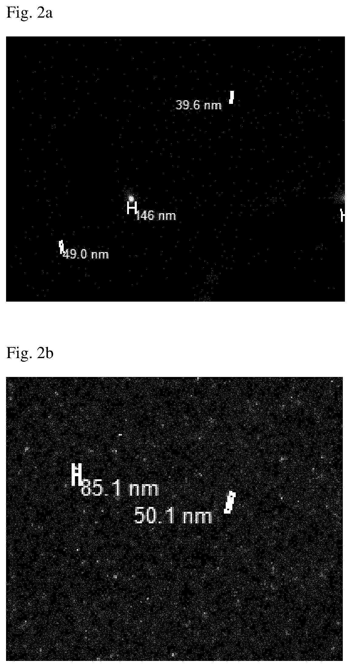
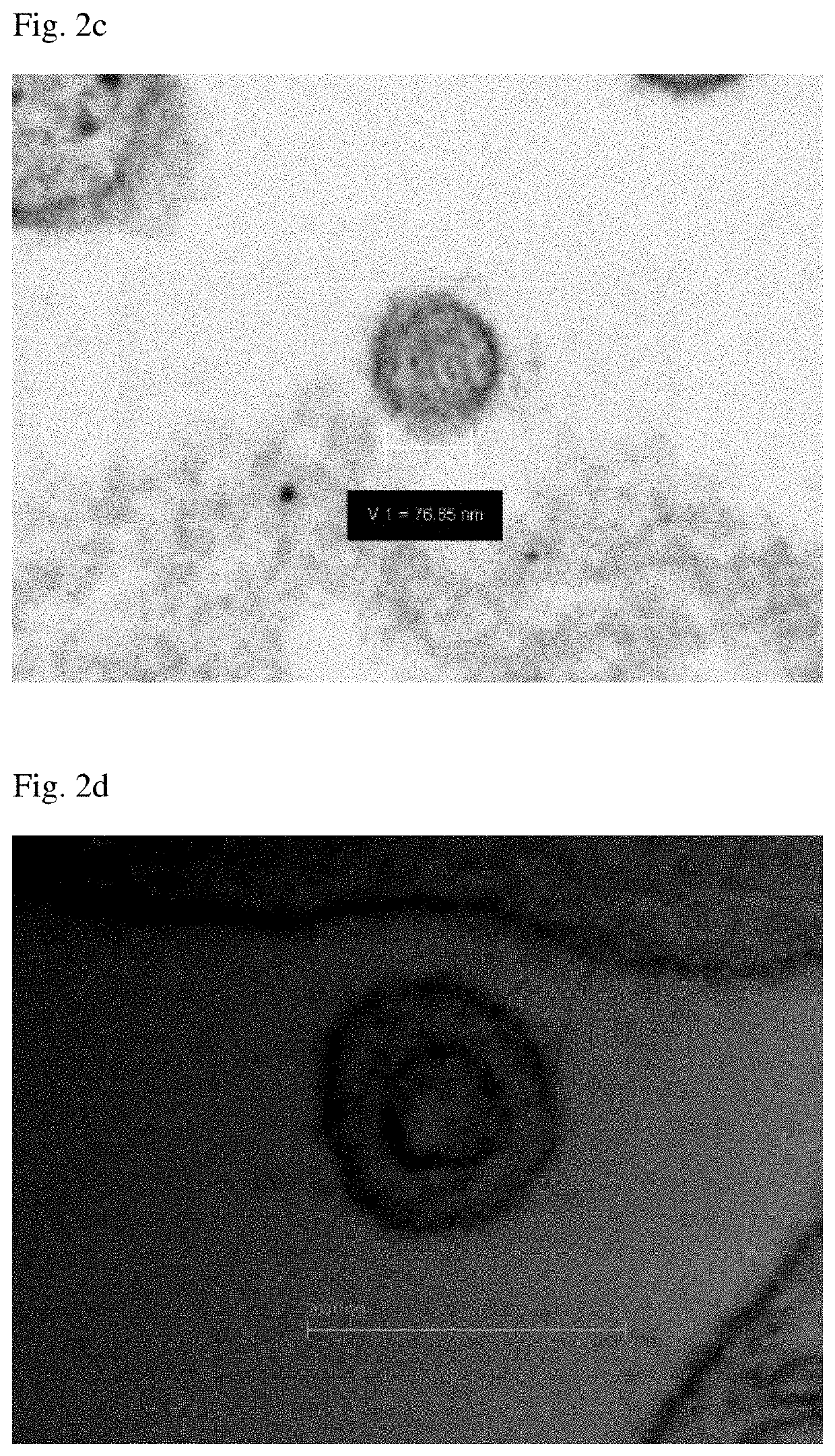
[0128](1) Structure
[0129]Among the luterials obtained in Example 1, luterials having a size of about 50-400 nm were imaged with a confocal laser scanning microscope (Zeiss), a transmission electron microscope, a scanning electron microscope, an atomic force microscope and a confocal scanner (Leica TCS-SP8). As a result, it was shown that the luterials also had a multiple ring-like layers of membrane structure and a non-completed internal cristae structure, similar to mitochondria, and were observed in the same wavelength range as that for mitochondria. In addition, it could be observed that the luterials were circular or oval in shape (FIGS. 1, 1(e), 2(h), 13 and 14).
[0130](2) Staining Characteristics
[0131]Among the luterials obtained in Example 1, luterials having a size of about 50-800 nm were stained with Mito-tracker, Rhodamine 123, Acridine Orange and Janus green B, and tested for their positive staining. The results showed that even the plant-derived luteria...
PUM
| Property | Measurement | Unit |
|---|---|---|
| size | aaaaa | aaaaa |
| size | aaaaa | aaaaa |
| size | aaaaa | aaaaa |
Abstract
Description
Claims
Application Information
 - R&D
- Intellectual Property
- Life Sciences
- Materials
- Tech Scout
- Unparalleled Data Quality
- Higher Quality Content
- 60% Fewer Hallucinations
Browse by: Latest US Patents, China's latest patents, Technical Efficacy Thesaurus, Application Domain, Technology Topic, Popular Technical Reports.
© 2025 PatSnap. All rights reserved.Legal|Privacy policy|Modern Slavery Act Transparency Statement|Sitemap|About US| Contact US: help@patsnap.com