Neuromodulation device
a neuromodulation device and neuromodulation technology, applied in contraceptive devices, artificial respiration, therapy, etc., can solve the problems of increased circulating catecholamines likely to have associated side effects, increased heart rate and blood pressure, and increased circulating catecholamines, so as to prevent or ameliorate bronchoconstriction, preserve neuronal structure and function, and minimal extrapulmonary side effects
- Summary
- Abstract
- Description
- Claims
- Application Information
AI Technical Summary
Benefits of technology
Problems solved by technology
Method used
Image
Examples
example 1
Ex Vivo Model of Bronchoconstriction
[0175]The methods for studying vagally-mediated bronchoconstriction have been described in detail elsewhere (Canning et al., Am J Physiol Regul Integr Comp Physiol. 2002 August; 283(2):R320-30). The airways and associated nerves are dissected free of all extraneous tissues and placed in water-jacketed dissecting dish continuously perfused with warmed, oxygenated Krebs buffer. A mainstem bronchus is isolated with associated nerves intact. Stirrups are placed on either side of the bronchus, with one fixed to the bottom of the recording chamber and the second attached to an isometric force transducer. The associated vagus nerves are stimulated (0.1-64 Hz) electrically using bipolar electrodes, resulting in muscle contraction.
[0176]In an isolated ex vivo guinea pig vagus-bronchus preparation, a low frequency electrical stimulation of the whole vagus nerve activates preganglionic_parasympathetic nerves lead to a rapid cholinergic contraction of the bro...
example 2
Ex-Vivo Assessment of KFAC on Compound Action Potential Conduction in Whole Vagus Nerve and Thoracic Branches
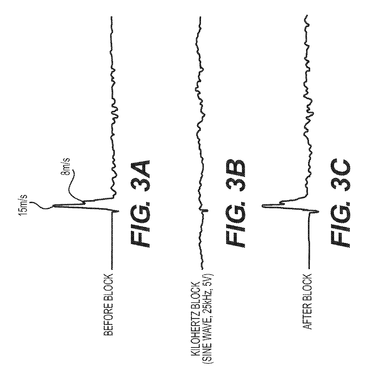
[0179]Vagus nerves obtained from guinea pigs and vagus thoracic branches obtained from human organ donors were dissected free from surrounding tissues. One end of the cut vagus nerve or branch was stimulated via suction electrodes attached to a stimulator that delivered single rectangular pulses. Compound action potentials were recorded at the other end of vagus nerve or thoracic branch nerve using a conventional recording suction electrode. The resulting signals were amplified (AM Systems, Model 1800), displayed on an oscilloscope and stored on a computer. During application of a neuromodulatory electrical signal between the stimulating and recording electrode, the amplitude of the waves in the compound action potential are reduced compared with the amplitude recorded prior to the application of the neuromodulatory signal (FIG. 8, Direct Current) and FIG. 9, Alternating Curr...
example 3
In Vivo Model of Bronchoconstriction
[0180]Methods for studying vagally-mediated bronchospasm in anesthetized guinea pigs have been described in detail elsewhere (Mazzone and Canning, Curr Protoc Pharmacol. 2002 May 1; Chapter 5:Unit 5.26; Auton Neurosci. 2002 Aug. 30; 99(2):91-101) and shown in FIG. 10. Guinea pigs are anesthetized with urethane (1.5 g / kg ip). The trachea and vagus nerves are visualized by a midline incision in the neck. The trachea is cannulated and connected to a constant volume ventilator (6 mL / kg body weight). The animals are then paralyzed with succinylcholine (2.5 mg / kg sc). An artery and vein are cannulated to monitor cardiovascular parameters and for drug delivery. The vagus nerves are placed on bipolar electrodes. A pressure transducer connected to a sideport of the tracheal cannula is used to monitor pulmonary inflation pressure. Bronchospasm is recorded as a percentage increase in pulmonary inflation pressure.
[0181]An in vivo guinea pig model of bronchoco...
PUM
 Abstract
Description
Claims
Application Information
 - R&D
- Intellectual Property
- Life Sciences
- Materials
- Tech Scout
- Unparalleled Data Quality
- Higher Quality Content
- 60% Fewer Hallucinations
Browse by: Latest US Patents, China's latest patents, Technical Efficacy Thesaurus, Application Domain, Technology Topic, Popular Technical Reports.
© 2025 PatSnap. All rights reserved.Legal|Privacy policy|Modern Slavery Act Transparency Statement|Sitemap|About US| Contact US: help@patsnap.com


