
Devices, kits and methods relating to treatment of facet joints

a facet joint and kit technology, applied in the field of spine and facet joints, can solve the problems of spinal instability, cysts along the back of the joints, surgery damage to surrounding tissues, etc., and achieve the effect of preventing tissue coring

Active Publication Date: 2019-03-26
THIXOS
View PDF102 Cites 0 Cited by
  • Summary
  • Abstract
  • Description
  • Claims
  • Application Information

AI Technical Summary

Benefits of technology

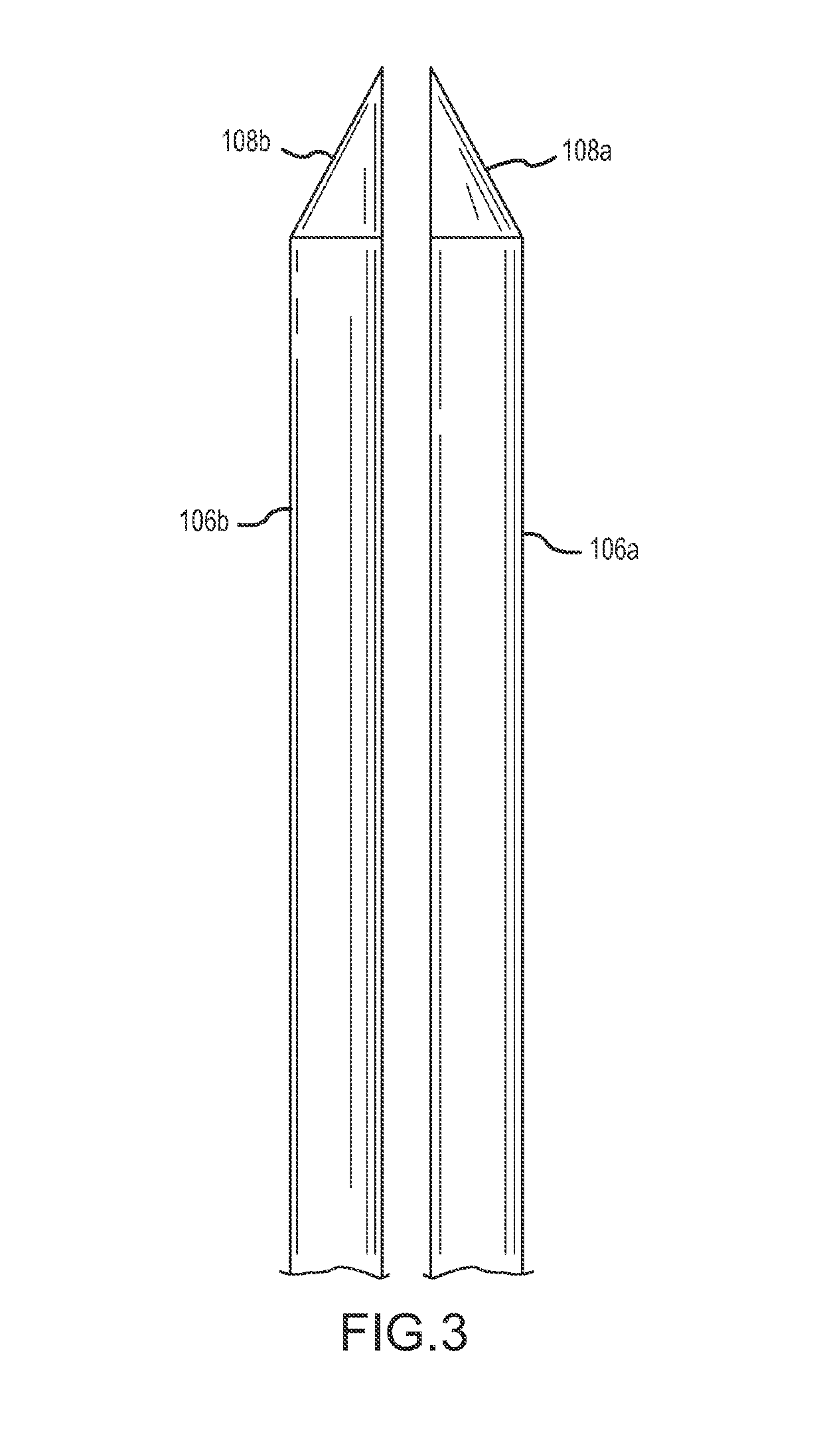
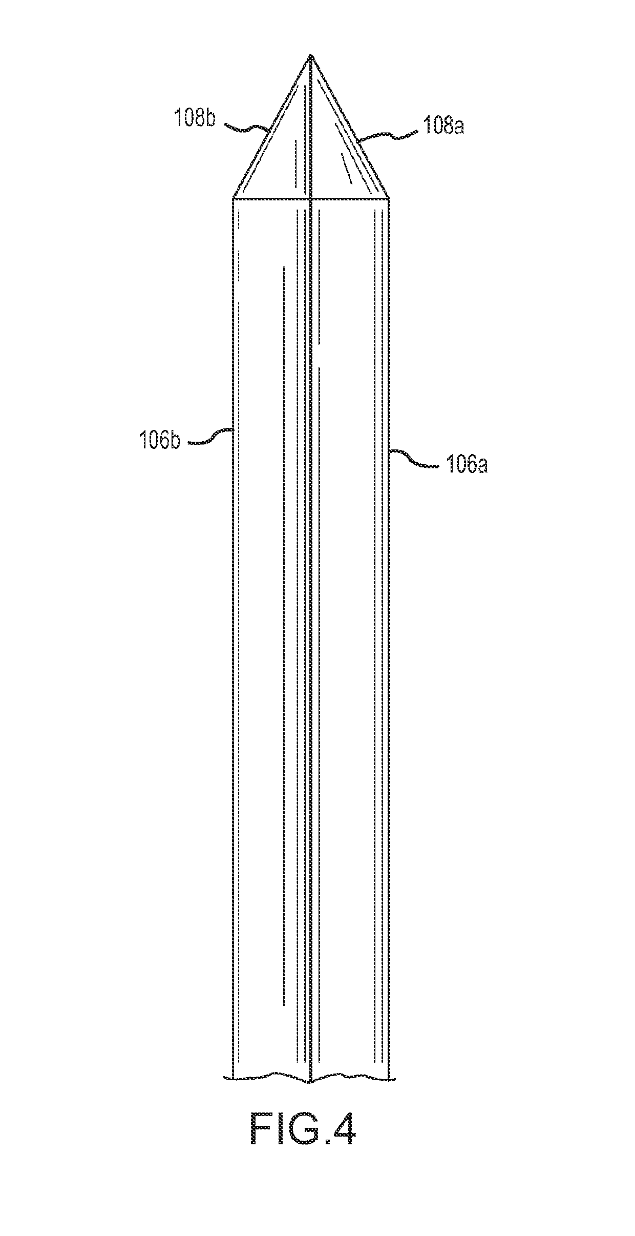
[0026]Such a stylet may have a beveled tip that corresponds with a beveled distal tip of the corresponding rotatable member. The stylet may be configured so that when fully inserted through the rotatable member, the distal tip of the stylet aligns with the distal tip of the rotatable member to form a uniform penetrating tip, with the distal end of the stylet blocking the distal end of the lumen to prevent coring of tissue.
[0032]The sheath may advantageously have a tapered distal end portion, wherein an exterior cross-section of the sheath tapers to a smaller size moving toward the distal end of the sheath. Such a tapered distal end portion of the sheath facilitates clean penetration of tissue when the facet joint surgical tool is configured to pierce through tissue to access a facet joint. The exterior cross-section taper may be accomplished through gradually reducing the wall thickness of the sheath on the distal end portion of the sheath, for example by grinding off a portion of the wall thickness from a portion of the sheath adjacent the distal end. In preferred implementations, an internal passage through the sheath does not taper to a smaller size in the tapered distal end portion of the sheath. Such a tapered distal end portion may extend from the distal end of the sheath to a location that may be at least 1 millimeter, at least 1.5 millimeters, at least 2 millimeters or at least 2.5 millimeters. Such a tapered distal end portion of the sheath may begin at a location not more than 10 millimeters, not more than 7 millimeters, not more than 5 millimeters, not more than 4 millimeters or not more than 3 millimeters from the distal end of the sheath.

Problems solved by technology

Cysts along the back of the joints may be associated with back pain but are usually asymptomatic.
However surgery damages surrounding tissues, ligaments and muscles.
Also surgery can result in disability due to removal of or damage to the muscle, ligaments and bony structures that mechanically support the spine and may result in spinal instability.
Although surgery is usually successful cysts may recur even after a successful operation.
Synovial fluid is very thick and often cannot be aspirated using a standard spinal needle.
Also, even when successfully aspirated the cysts may refill with joint fluid resulting in recurrence of symptoms.
Therefore many percutaneous aspirations fail to resolve the patient's symptoms and patients often have to undergo a second open surgical procedure to definitively treat their pain with additional risks, expense, recovery time and pain.
Direct percutaneous aspiration has traditionally not been an option for synovial cysts located anterior to facet joints.
The nerves supplying the facet joint are heated and this disrupts the nerve function and conduction resulting in pain relief.

Method used


Image

  • Devices, kits and methods relating to treatment of facet joints
  • Devices, kits and methods relating to treatment of facet joints
  • Devices, kits and methods relating to treatment of facet joints

Examples


Embodiment Construction

[0068]For a more complete understanding of the invention and aspects thereof, reference is now made to the following Detailed Description taken in conjunction with the figures.

[0069]In one embodiment, an access device is formed by two elongated round hollow metal tubes of circular cross-section. The ends of the tubes may be cut at an angle. The tube edges may be sharpened, such as a pointed tip of a spinal needle or hypodermic needle. The two tubes may be inserted through and constrained by a smooth outer sheath. Thus the outer sheath may be oval shaped in cross section and rigid. The rigid sheath may prevent the tubes from crossing or overlapping during insertion or rotation of the tubes. The sheath may be sized to allow the tubes to freely rotate. Before the access device is inserted into the skin and deep tissues, the angled ends of the tubes may first be rotated to face in opposite directions. The two sharpened edges may now form a single blade-like surface. For convenience such...


PUM


Abstract

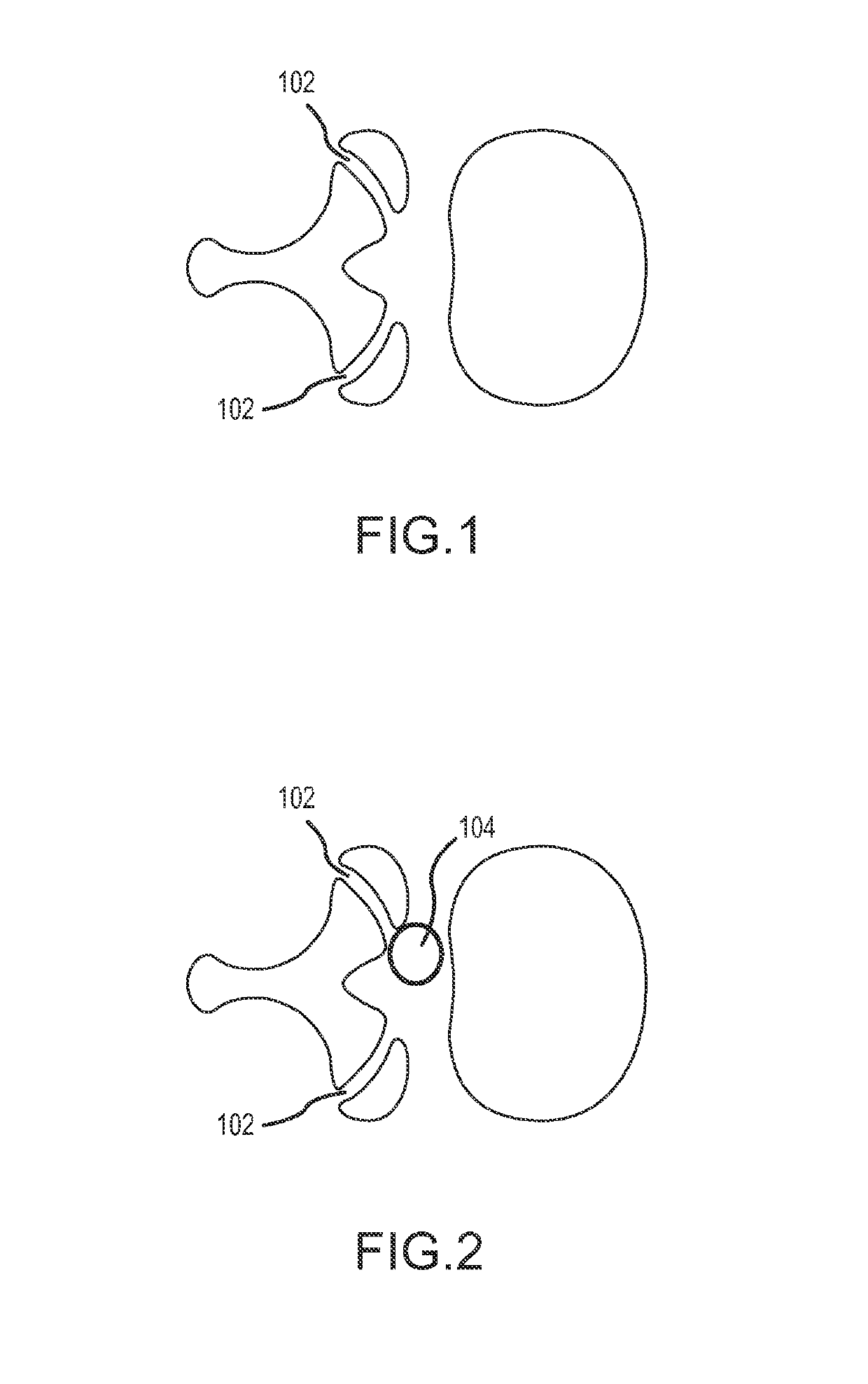
A facet joint surgical tool for treating a facet joint synovial cyst includes rotatable members disposed side-by-side through a minimally invasive sheath and rotatable to reconfigure distal end portions between a facet joint penetration configuration with a tissue piercing tip and a facet joint retraction configuration. Facet joint synovial cysts located to an anterior side of the facet joint are treated by a posterior approach with access to the cyst through the facet joint retracted by the surgical tool. Facet joint synovial cysts located to a posterior side of the facet joint are treated by direct access from a posterior approach.

Description

CROSS-REFERENCE TO RELATED APPLICATIONS[0001]This application claims a benefit of U.S. provisional patent application No. 61 / 903,806 entitled “DEVICES, KITS AND METHODS RELATING TO TREATMENT OF FACET JOINTS” filed Nov. 13, 2013, each and every portion of which is incorporated herein by reference.FIELD OF INVENTION[0002]This invention relates to the spine and facet joints, including with respect to minimally invasive procedures to treat facet joint synovial cysts, which may be image guided.BACKGROUND OF INVENTION[0003]Back pain is the commonest cause of disability. Approximately 40% of back pain is caused by disorders of the facet joints.[0004]The facet joints (also called zygapophysial joints or z-joints) are paired structures on the back and side of the spine. These joints support the torso during flexion, extension and rotation of the spine. These are synovial joints and therefore contain joint fluid. Joint fluid is viscous and a lubricant. Synovial cysts occur along the margins o...

Claims


Application Information

Patent Timeline
Patent Type & Authority Patents(United States)
IPC IPC(8): A61B1/32A61B18/14A61B18/12A61B17/34A61B17/02A61B18/00A61B17/32
CPCA61B18/1482A61B17/3417A61B17/3421A61B18/1206A61B17/3439A61B18/1487A61B2217/007A61B2017/320044A61B2017/3454A61B2018/00339A61B2018/00577A61B2217/005A61B2017/0256
Inventor SOLSBERG, MURRAY DAVIDBEALL, DOUGLAS PRESTONDEAN, RYAN ERICH
Owner THIXOS
Learn More