Preparation method and application of red light carbon dots and micelles
A technology of carbon dots and red light, which is applied in the field of preparation of carbon dots and micelles, can solve the problems of multiple cleanings, short emission wavelengths, complex purification steps, etc., and achieve the effects of avoiding interference, enhancing water dispersibility, and low cost
- Summary
- Abstract
- Description
- Claims
- Application Information
AI Technical Summary
Problems solved by technology
Method used
Image
Examples
Embodiment 1
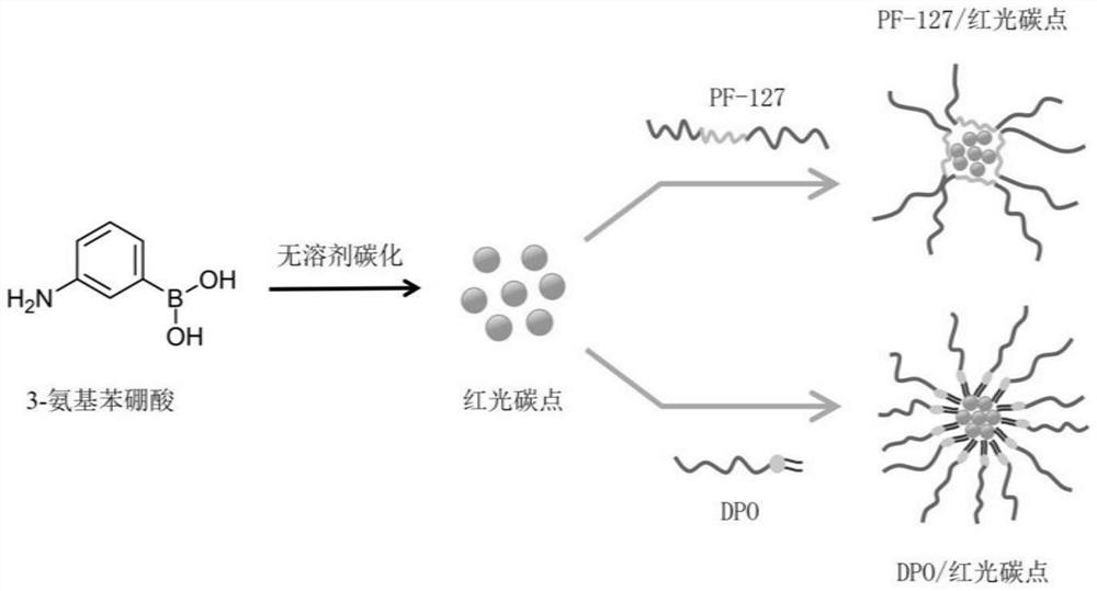
[0027] The preparation steps of the red light carbon dots in this embodiment are as follows:
[0028] (1) Weigh 1g of 3-aminophenylboronic acid solid powder and add it to a 10mL polytetrafluoroethylene lining, and put the lining into a matching autoclave;
[0029] (2) Reaction: the autoclave is sealed and reacted in an oven at 220° C. for 20 h, then taken out to room temperature and cooled naturally;
[0030] (3) Purification: The product was washed three times with deionized water to remove impurities, and then the obtained solid was placed in an oven at 60° C. and dried overnight to obtain the final product (ie, red light carbon dots), which was stored in a refrigerator at 4° C.
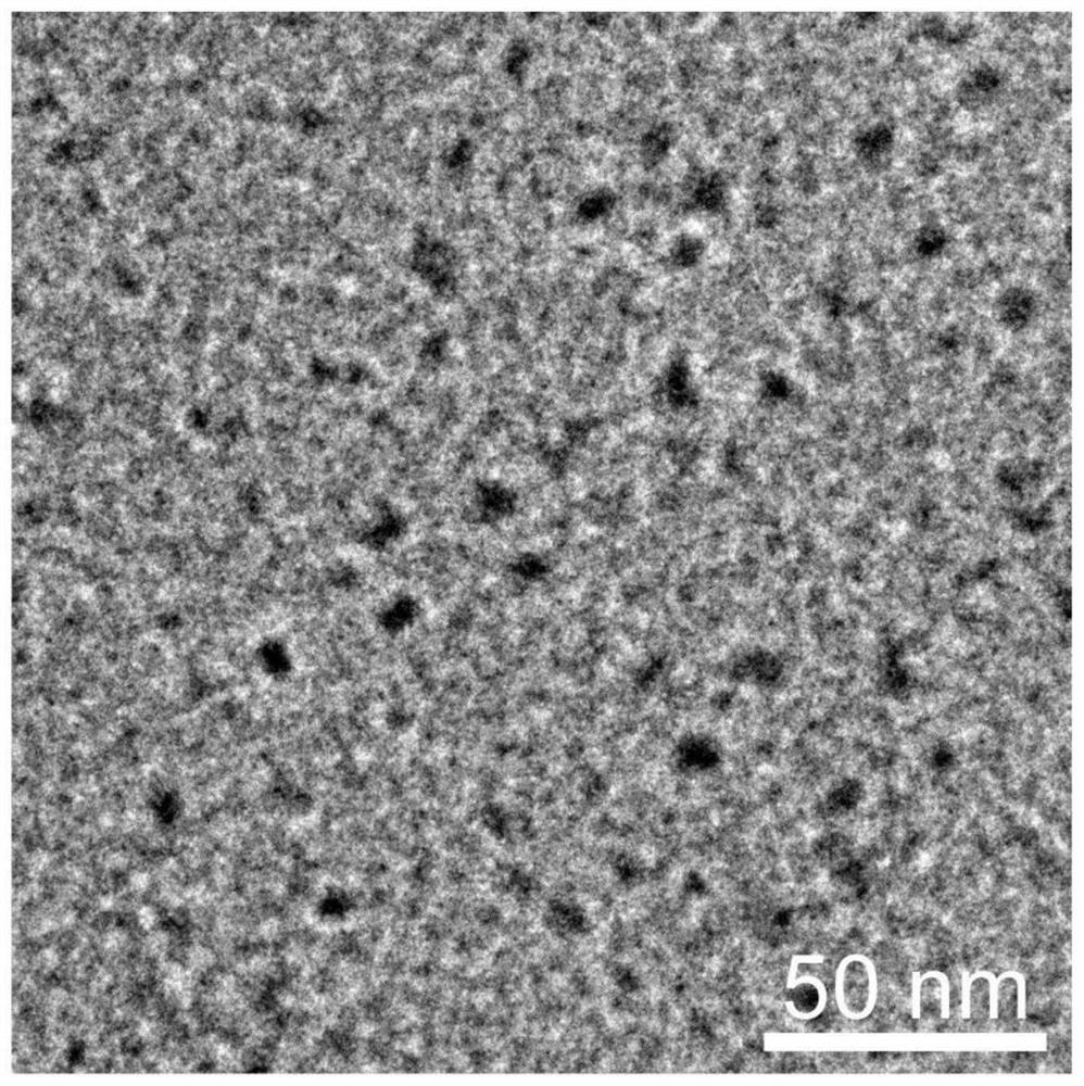
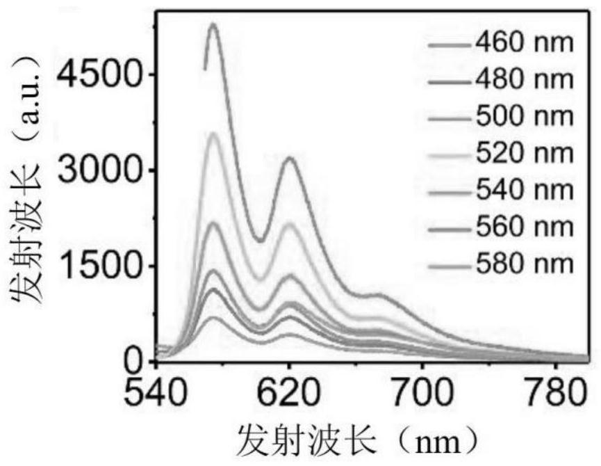
[0031] The red light carbon dots obtained by the above method are dispersed in ethanol solvent and the transmission electron microscope picture obtained is shown in figure 2 . Experimental results show that the particle size of the red carbon dots is 6.9nm. After the red carbon dots obtained by...
Embodiment 2
[0033] To test the imaging effect of the red light carbon dots prepared in Example 1 on the lipid droplets of A549 cells, the method is as follows:
[0034] (1) Cell culture: A549 cells were cultured at 5×10 4 Seed in a 96-well plate at a density of 1 / mL (100 μL) at 37°C, 5% CO 2 Cultivate in the environment for 24h;
[0035] (2) Cell staining: wash three times with phosphate buffered saline (PBS), add 100 μL PBS containing 20 μg / mL red light carbon dots and commercial lipid droplet fluorescent dye BODIPY 493 / 503 (1 μg / mL) and incubate for 15 min, wash with PBS cells 3 times;
[0036] (3) Confocal fluorescence microscopy imaging observation: Confocal fluorescence images were taken under the excitation of 552nm laser (red light carbon dots) and 488nm laser (BODIPY 493 / 503) using a laser confocal fluorescence microscope.
[0037] The results of confocal fluorescence imaging of cells after co-staining of red carbon dots and BODIPY 493 / 503 observed by the above method are as fo...
Embodiment 3
[0039] The preparation of micelles based on red light carbon dots in this embodiment, the specific steps are as follows:
[0040] (1) Preparation: Disperse 20mg of PF-127 or DPO and 2mg of red carbon dots in 0.5mL of dichloromethane, and dry with nitrogen;
[0041] (2) Purification: drying the sample obtained above in a vacuum drying oven at room temperature for more than 1 h;
[0042] (3) Resuspension: Add 1 mL of deionized water to each dried sample, and sonicate the resulting suspension at 25°C for 30 min to prepare a lipid droplet-specific targeting gel based on red light carbon dots. bundles (i.e. "PF-127 / Red Carbon Dots" and "DPO / Red Carbon Dots").
[0043] The transmission electron microscope pictures of lipid droplet-specific targeting micelles based on red light carbon dots obtained by the above method are shown in Figure 5 . The results show that the micelles based on red light carbon dots have good water dispersibility and uniform particle size. The particle size ...
PUM
 Abstract
Description
Claims
Application Information
 - R&D
- Intellectual Property
- Life Sciences
- Materials
- Tech Scout
- Unparalleled Data Quality
- Higher Quality Content
- 60% Fewer Hallucinations
Browse by: Latest US Patents, China's latest patents, Technical Efficacy Thesaurus, Application Domain, Technology Topic, Popular Technical Reports.
© 2025 PatSnap. All rights reserved.Legal|Privacy policy|Modern Slavery Act Transparency Statement|Sitemap|About US| Contact US: help@patsnap.com


