Nocardia cardia bacteriophage P3.2 and application thereof
A Nocardia and phage technology, applied in the fields of phage, virus/phage, biological water/sewage treatment, etc., can solve the problems of water deterioration, difficult to remove foam, etc., to improve the breadth and effectiveness, expand the host spectrum, Fast and efficient cracking effect
- Summary
- Abstract
- Description
- Claims
- Application Information
AI Technical Summary
Problems solved by technology
Method used
Image
Examples
Embodiment 1
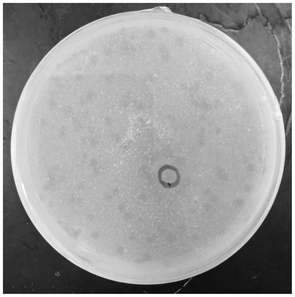
[0019] The separation and purification of embodiment 1 phage
[0020] Preparation of PYCa liquid medium:
[0021] at 1L ddH 2 Add 1 g of glucose, 1 g of yeast extract, and 15 g of tryptone to O, mix well and then autoclave at 121°C for 20 min. After cooling, add 4.5 mL of calcium chloride solution with a concentration of 1 mol / L.
[0022] Preparation of PYCa solid medium:
[0023] at 1L ddH 2 Add 1g of glucose, 1g of yeast extract, 15g of tryptone, and 15g of agar powder to O, mix well and then autoclave at 121°C for 20min. Add 4.5mL calcium chloride solution with a concentration of 1mol / L in the uncoagulated state.
[0024] Preparation of PYCa semi-solid medium:
[0025] at 1L ddH 2 Add 1 g of glucose, 1 g of yeast extract, 15 g of tryptone, and 7.5 g of agar powder into O, mix well, and then sterilize under high pressure at 121 ° C for 20 min. Add 4.5mL calcium chloride solution with a concentration of 1mol / L in the uncoagulated state.
[0026] The sewage sample use...
Embodiment 2
[0029] Morphological observation of embodiment 2 bacteriophage
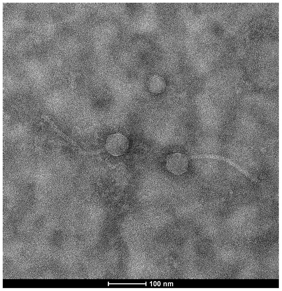
[0030] Take 20 μL of the phage suspension and drop it on the copper grid, wait for its natural precipitation for 10 minutes, blot it dry from the side with dry filter paper, let it air for about 1 minute, add 1 drop of 1% uranyl acetate on the copper grid, stain for 2 minutes, and then use it carefully to dry Blot the excess dye from the side of the filter paper, let it dry naturally in the dark for 30 minutes, and observe it with a transmission electron microscope (JEM-1400).
[0031] The results of transmission electron microscopy are shown in figure 2 , the results showed that bacteriophage P3.2 had a spherical head and a long tail. The length of the P3.2 tail was about 197.49nm, the diameter of the head was about 56.69nm, and the diameter of the tail was about 14.57nm. According to the eighth report on the virus classification of the International Organization for Taxonomy of Viruses (ICTV), this strain of ...
Embodiment 3
[0032] Example 3 Phage Genome Analysis and Identification
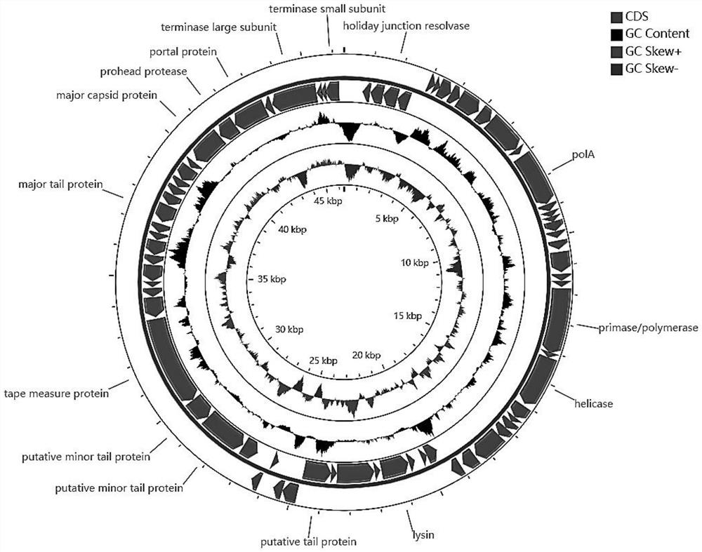
[0033] Phage whole genome sequencing and analysis: IlluminaNextSeq500 was used for PE 2×150 to sequence the DNA of the sample. Then, the optimized sequence was spliced using spades v.3.11.1 splicing software, and the optimal assembly result was obtained. Use the biological software PHASTER to predict and analyze the open reading frame of the genome, and use NCBI Blastp to complete the preliminary annotation of functional genes, and use tRNAscan-SE (http: / / lowelab.ucsc.edu / / tRNAscan-SE / ) to predict tRNA online, Use CGView Server software (http: / / cgview.ca / ) to complete the drawing of the whole genome circle map (see image 3 ), a phylogenetic tree was constructed using MEGA X software based on the whole genome data (see Figure 4 ).
[0034]The genome size of phage P3.2 is 46380bp, and the G+C content is 67.1mol%. According to the results of Blast comparison, it is also a new phage, and the most homologous to it ...
PUM
| Property | Measurement | Unit |
|---|---|---|
| diameter | aaaaa | aaaaa |
Abstract
Description
Claims
Application Information
 - R&D
- Intellectual Property
- Life Sciences
- Materials
- Tech Scout
- Unparalleled Data Quality
- Higher Quality Content
- 60% Fewer Hallucinations
Browse by: Latest US Patents, China's latest patents, Technical Efficacy Thesaurus, Application Domain, Technology Topic, Popular Technical Reports.
© 2025 PatSnap. All rights reserved.Legal|Privacy policy|Modern Slavery Act Transparency Statement|Sitemap|About US| Contact US: help@patsnap.com


