CBCT image artifact removing method

A CT image and artifact removal technology, applied in the field of image processing, can solve the problems of noise and artifacts, image quality degradation, low soft tissue contrast, etc., and achieve the effect of not easily deformed, high resolution, and easy to observe

Pending Publication Date: 2022-02-01
NANJING UNIV OF POSTS & TELECOMM
View PDF0 Cites 0 Cited by
  • Summary
  • Abstract
  • Description
  • Claims
  • Application Information

AI Technical Summary

Problems solved by technology

[0005] Compared with conventional fan-beam CT images, the image quality of CBCT is degraded due to x-ray scatter and truncated projections, and suffers from low soft tissue contrast, noise, and artifacts, which hinder the effective use of CBCT in many potential applications

Method used


Image

  • CBCT image artifact removing method
  • CBCT image artifact removing method
  • CBCT image artifact removing method

Examples


Embodiment 1

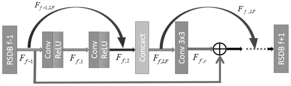
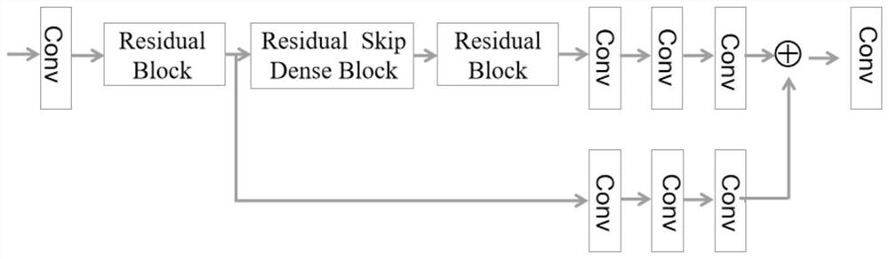
[0052] Such as figure 1 As shown, a CBCT image de-artifact method based on contextual loss and feature fusion residual network disclosed in this embodiment specifically includes the following steps:

[0053] Step (1) preprocesses each image in the CBCT and CT datasets so that each image has the same size and is a suitable input format for the network. Specifically include the following sub-steps:

[0054] (1.1) The CBCT and CT data (.dcm format) obtained from the hospital are intercepted to the same size, the pixel size is m*n, m and n are the length and height of the image respectively, and m=n=512 is taken in the experiment;

[0055] (1.2) Convert the resized .dcm data into .raw format data suitable for the network.

[0056] The data used in this example comes from a hospital, and then we make data sets for different parts, including chest, head and pelvis. In practice, it can also be adapted to other parts. Preprocess these data, intercept each image to 512*512 pixels, ...


PUM


Abstract

The invention discloses a CBCT image artifact removing method. Based on contextual loss and a feature fusion residual network, the method comprises the following steps firstly, performing feature extraction on an input CBCT image and an input CT image with artifacts to obtain a feature spectrum; performing improved feature fusion residual network training by taking context loss as a loss function on the obtained feature map, learning the structural similarity between CBCT and CT images, and taking the CT images as labels, so that artifacts of the CBCT images can be removed while the structures of the CBCT images are kept unchanged; and finally, taking the CBCT image as input, and performing artifact removal by utilizing the trained network model. According to the method, non-alignment between input images can be allowed by using the contextual loss, the method can be well suitable for the characteristic that CBCT and CT medical images cannot be strictly aligned, and finally, the effect of rapidly and effectively removing artifacts is achieved.

Description

technical field [0001] The invention relates to a method for removing artifacts of a CBCT image, in particular to a method for removing artifacts of a CBCT image based on contextual loss and a feature fusion residual network, and belongs to the technical field of image processing. Background technique [0002] Cone-beam computed tomography (CBCT) has the advantages of short scanning time and low X-ray dose, and is widely used in the diagnosis of head and neck diseases. However, compared to conventional fan-beam computed tomography (CT), CBCT images suffer from reduced quality due to X-ray scattering and projection truncation. These problems lead to large scattering artifacts, which affect the application prospect of CBCT in many fields. Therefore, the main challenge for CBCT is scattering artifacts. [0003] In recent years, many researchers have studied CBCT scatter correction. In the published literature, the main correction methods can be divided into two categories ac...

Claims


Application Information

Patent Timeline
IPC IPC(8): G06T11/00G06V10/74G06V10/80G06V10/82G06K9/62G06N3/04G06N3/08
CPCG06T11/003G06N3/08G06T2211/424G06N3/045G06F18/22G06F18/253
Inventor 谢世朋严墨
Owner NANJING UNIV OF POSTS & TELECOMM
Why Eureka
Try Eureka