Cross-species cleavable xanthomonas phage as well as composition, kit and application thereof
A technology of Xanthomonas and phage, applied in the field of phage, can solve the problems of little research and achieve the effect of high specificity and lysis, high affinity and lysis ability, and wide host range
- Summary
- Abstract
- Description
- Claims
- Application Information
AI Technical Summary
Problems solved by technology
Method used
Image
Examples
Embodiment 1
[0062] Example 1 Isolation, preparation and purification of Xanthomonas bacteriophage GJ19P1 The source samples of Xanthomonas bacteriophage GJ19P1 in the present invention were collected from sewage in Jiangning District, Nanjing City, Jiangsu Province, filtered through double-layer filter paper, centrifuged at low speed and normal temperature, and then 0.22 Filter the supernatant through a μm filter membrane.
[0063] Phage Isolation:
[0064] (1) Take 10 mL of the filtered supernatant, add it to 10 mL of 2 times TSB liquid medium, and add 1 mL of phage host bacteria GJ19 logarithmic phase bacterial liquid at the same time, and place it at 28 ° C for overnight culture;
[0065] (2) Take the above culture, centrifuge at 8000rpm for 10min, filter the supernatant with a 0.22μm filter membrane, and set aside;
[0066] (3) Take 0.5 mL of the bacteriophage host bacteria Xanthomonas rugosa GJ19 in the logarithmic phase, add 5 mL, 40 ° C TSB semi-solid agar medium, mix well, pour i...
Embodiment 2
[0075] Example 2 Electron Microscopic Observation of Xanthomonas Phage GJ19P1
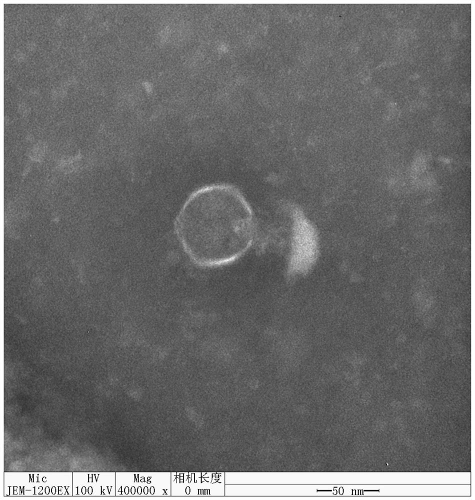
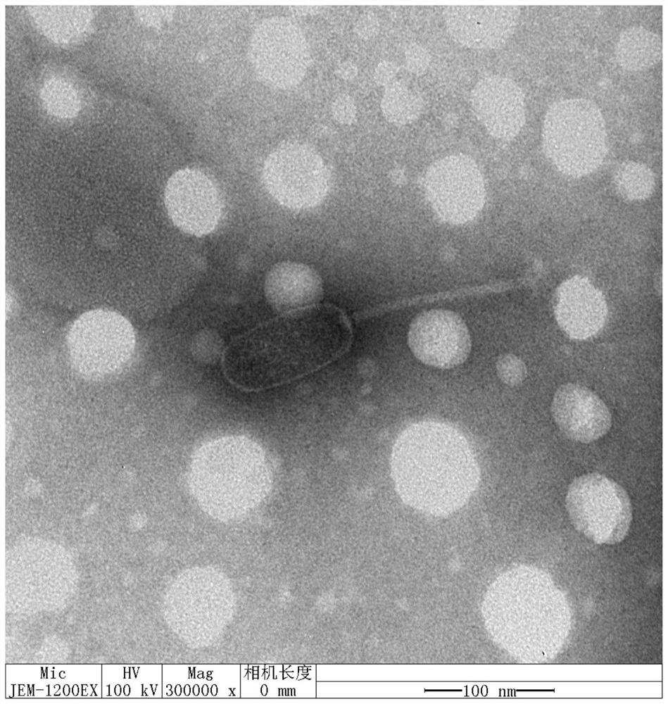
[0076] Take the purified phage solution prepared in Example 1 for electron microscope observation: take 20 μL of the sample and drop it on the copper grid, wait for its natural precipitation for 15 minutes, absorb the excess liquid from the side with filter paper, add 1 drop of 2% phosphotungstic acid on the copper grid, and stain for 10 minutes , Use filter paper to absorb the dye solution from the side, and observe it with an electron microscope after drying.
[0077] The result is as figure 2 As shown, observing the morphology of Xanthomonas phage GJ19P1 under an electron microscope reveals that the phage presents a polyhedral stereosymmetric head with a diameter of about 60 nm and a short contracted tail under an electron microscope. The phage belongs to the Autographiviridae family of self-replicating short-tailed bacteriophages.
Embodiment 3
[0078] Example 3 Preparation of Xanthomonas bacteriophage GJ19P1 particles and extraction and sequencing of genome
[0079] (1) Take 100 mL of the purified phage solution prepared in Example 1, add 20 μL of DNaseI and 20 μL of RNaseA at a concentration of 5 mg / mL in sequence, incubate at 37°C for 60 minutes, add 5.84 g of NaCl, and place in an ice bath for 1 hour after dissolution;
[0080] (2) Centrifuge at 11,000rpm for 10min at 4°C, transfer the centrifuged supernatant to a new centrifuge tube, add solid PEG8000 to make the final concentration 10% (w / v), and after the PEG8000 is completely dissolved, Ice bath for 1h;
[0081] (3) Centrifuge at 11,000 rpm for 20 minutes at 4°C, add 1 mL of SM solution to resuspend the precipitate, and obtain a phage particle concentrate, which is stored at 4°C until use.
[0082] Phage nucleic acid was extracted and sequenced using λ phage genomic DNA kit. After nucleotide sequencing, the Xanthomonas phage GJ19P1 (Xanthomonas phage PSA-P1)...
PUM
| Property | Measurement | Unit |
|---|---|---|
| diameter | aaaaa | aaaaa |
Abstract
Description
Claims
Application Information
 - R&D
- Intellectual Property
- Life Sciences
- Materials
- Tech Scout
- Unparalleled Data Quality
- Higher Quality Content
- 60% Fewer Hallucinations
Browse by: Latest US Patents, China's latest patents, Technical Efficacy Thesaurus, Application Domain, Technology Topic, Popular Technical Reports.
© 2025 PatSnap. All rights reserved.Legal|Privacy policy|Modern Slavery Act Transparency Statement|Sitemap|About US| Contact US: help@patsnap.com


