Salmonella enteritidis bacteriophage and application thereof
A technology of Salmonella enteritidis and Salmonella, applied in the direction of phage, virus/phage, application, etc., can solve the problems that limit the development and application of phage therapy, achieve the effect of broadening the range of lysis and application, and enriching the phage species library
- Summary
- Abstract
- Description
- Claims
- Application Information
AI Technical Summary
Problems solved by technology
Method used
Image
Examples
Embodiment 1
[0015] Embodiment 1, the separation and purification of phage
[0016] In this experiment, plastic bottles sterilized by high pressure steam were used, and the Dongjiang river water in Dongguan was collected as a sample. The water sample was centrifuged at 8000r / min for 15min, and the supernatant was filtered with a 0.22um filter syringe. The frozen Salmonella Enteritidis (GIM1.1105) bacterial liquid was inoculated in the chromogenic medium in advance, placed in a 37°C incubator for about 12 hours, and a single colony was inoculated into the LB liquid medium, 37°C, 180r / min shaking culture to the logarithmic phase to obtain fresh bacterial liquid. Take 5 mL of the filtered supernatant and put it into 20 mL of LB liquid medium, add 500 μL of bacterial liquid and 2 mL of SM buffer, mix well, and then place it on a shaker at 37°C and 220 r / min for 12 hours to obtain the enrichment solution. The enriched solution was centrifuged and filtered repeatedly to obtain the phage supern...
Embodiment 2
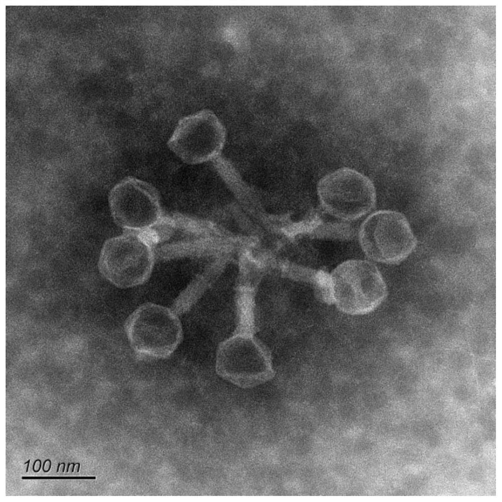
[0017] Embodiment 2, electron microscope observation of phage
[0018] Take 10 μL of the phage filtrate after amplified culture and drop it on the copper grid, let it settle naturally for 10 minutes, blot the excess liquid from the side with filter paper, then add 1 drop of 2% phosphotungstic acid to the copper grid, stain for 10 min, and use it after the copper grid is dry. The morphology of PSM6 was observed by transmission electron microscope.
[0019] Electron microscope observation results show that the phage PSM6 has an icosahedral head and a retractable tail, which belongs to the muscle tail phage. The electron microscope photos show that the diameter of the head of PSM6 is about 69±5nm, and the length of the tail is about 115±6nm. For details, see figure 2 .
Embodiment 3
[0020] Embodiment 3, the mensuration of phage thermostability
[0021] Take 1000 μL phage filtrate (about 10 8 PFU / mL) in a 1.5ml EP tube, react in water baths at 40°C, 50°C, 60°C, 70°C, and 80°C for 2 hours, take samples every 20 minutes, and immediately place the samples on ice to cool, after 10 times After serial dilution, the titer of phage was determined, the survival of phage was analyzed, and the data were processed using GraphPad Prism 8.
[0022] The results of thermostability test showed that the titer of PSM6 decreased faster as the temperature increased. At 40°C for 2 hours, the phage PSM6 maintained its original activity and its titer basically remained unchanged. After acting at 60°C for 2 hours, the titer is still 10 7.4 PFU / mL, the survival rate is about 25%; at 80°C for 2 hours, the titer of PSM6 is the lowest, down to 10 6.2 PFU / mL. On the whole, the thermal stability of PSM6 is relatively strong, see image 3 .
PUM
| Property | Measurement | Unit |
|---|---|---|
| diameter | aaaaa | aaaaa |
Abstract
Description
Claims
Application Information
 - R&D
- Intellectual Property
- Life Sciences
- Materials
- Tech Scout
- Unparalleled Data Quality
- Higher Quality Content
- 60% Fewer Hallucinations
Browse by: Latest US Patents, China's latest patents, Technical Efficacy Thesaurus, Application Domain, Technology Topic, Popular Technical Reports.
© 2025 PatSnap. All rights reserved.Legal|Privacy policy|Modern Slavery Act Transparency Statement|Sitemap|About US| Contact US: help@patsnap.com


