CT ultra-low dose automatic three-dimensional positioning scanning method and system

A technology of three-dimensional positioning and scanning method, which is applied in the field of CT scanning, which can solve the problems of inaccurate measurement of the horizontal position/size of objects, increase the scanning time, and increase the patient dose, etc., so as to reduce the preparation time for scanning switching and eliminate noise artifacts , the effect of easy operation

Pending Publication Date: 2020-10-30
NANJING ANKE MEDICAL TECH CO LTD
View PDF12 Cites 2 Cited by
  • Summary
  • Abstract
  • Description
  • Claims
  • Application Information

AI Technical Summary

Problems solved by technology

Moreover, if two or more positioning images are required, the tube needs to be rotated to the designated position, and multiple positioning images are scanned by entering and exiting the bed multiple times, which will increase the scanning time and increase the patient dose
[0004] 2. Traditional methods cannot accurately locate the space
[0005] like figure 2 As shown, the same object will get different detector outputs at different heights, resulting in inaccurate geometric positioning, so the horizontal position / size of the object cannot be accurately measured on the positioning image
[0006] 3. The traditional method needs to manually select the scanning area
After the positioning image is collected, since there are only two-dimensional positioning images and only approximate spatial information can be provided, the operator can only estimate and scan the scanning area of ​​the organ

Method used


Image

  • CT ultra-low dose automatic three-dimensional positioning scanning method and system
  • CT ultra-low dose automatic three-dimensional positioning scanning method and system
  • CT ultra-low dose automatic three-dimensional positioning scanning method and system

Examples


Embodiment Construction

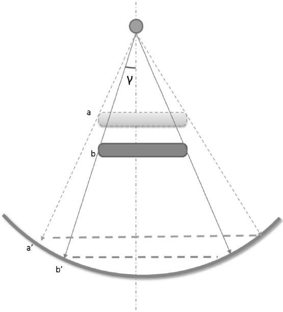
[0056] Such as Figure 1 to Figure 5 As shown, the present invention proposes a CT ultra-low-dose automatic three-dimensional positioning method in order to reduce scanning and scan preparation time and obtain scanning positioning information automatically and accurately under the same scanning dose.

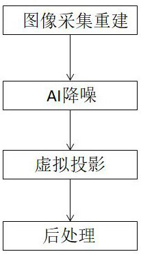
[0057] Such as image 3 As shown, the specific steps are as follows:

[0058] 1. Set the protocol parameters: set the scanning range of the 3D positioning image and the CT scanning protocol. The scanning of the 3D positioning image mainly refers to the starting position and the ending position of the movement of the bed. The CT scanning protocol includes the CT scanning position, scanning voltage and current etc., as well as the reconstructed layer thickness and layer spacing, etc.; the size and center of the reconstructed area usually need to be automatically generated according to the results of the scout scan;

[0059] 2. On the premise of keeping the frame rotating, the sc...


PUM


Abstract

The invention discloses a CT ultralow-dose automatic three-dimensional positioning scanning method and a system. The method comprises the following steps: under the condition of conventional radiationdose, scanning to obtain a normal dose image, adding quantum noise and electronic noise into the normal dose image to generate an ultralow dose spiral scanning image, and performing AI low dose reconstruction to obtain a three-dimensional positioning image for CT scanning. According to the invention, under the conditions of not stopping the rotation of the rack and adopting similar radiation doselevels, the three-dimensional positioning image is directly obtained, the area needing to be scanned is automatically positioned, automatic scanning is carried out, the positioning accuracy and scanning efficiency are improved, meanwhile, the radiation dose of a patient can be reduced, and the requirements for the technical level, experience and the like of scanning operators are reduced.

Description

technical field [0001] The invention relates to the technical field of CT scanning, in particular to a CT ultra-low dose automatic three-dimensional positioning scanning method and system capable of rapidly improving scanning efficiency. Background technique [0002] Before scanning, the existing CT (Computer Tomography, X-ray computer tomography system) usually needs to scan one or two positioning images to locate the scanned object and set the scanning range, such as the length of the scan, the location of the hospital bed, etc. Height, etc., according to the positioning image, determine the scanning range, angle, scanning parameters, delay time, etc. for the subsequent helical or axial scan. When scanning the positioning image, the tube that generates X-rays is usually fixed above or on the side of the patient to scan the patient. The main shortcomings of the existing positioning image technology are reflected in the following aspects: [0003] 1. It takes a long prepar...

Claims


Application Information

Patent Timeline
Patent Type & Authority Applications(China)
IPC IPC(8): A61B6/03A61B6/00G06T17/00G06N3/04G06N3/08G06F30/20G06F111/10
CPCA61B6/032A61B6/5211A61B6/5258A61B6/4411A61B6/54G06T17/00G06N3/08G06F30/20G06F2111/10G06N3/045
Inventor 曾凯冯亚崇吴小页
Owner NANJING ANKE MEDICAL TECH CO LTD
Why Eureka
Try Eureka