A method for establishing a sheep endometrial epithelial cell line and its application
A technology of endometrium and epithelial cells, applied in the biological field, can solve problems such as abnormal expression of target genes, silencing of vectors and foreign genes, and influence on the physiological state of cells, and achieve low nutritional requirements, shortened cell passage time, and high efficiency. The effect obtained
- Summary
- Abstract
- Description
- Claims
- Application Information
AI Technical Summary
Problems solved by technology
Method used
Image
Examples
Embodiment 1
[0042] Example 1 Establishment of Sheep Endometrium Epithelial Cell Line
[0043] 1. Collect sheep uterine horns, separate and culture endometrial epithelial cells by tissue block culture method, at 37 ℃, 5% CO 2 Cultivate in the incubator for 7-12 days.
[0044] 2. Using the liposome transfection method, the universal PB transposon carrier pTP-hTERT carrying the hTERT gene was introduced into the primary cultured sheep endometrial epithelial cells for transfection. The specific steps are as follows:
[0045] 1) Before transfection, inoculate the third-generation primary cultured sheep endometrial epithelial cells in a 24-well plate, and perform transfection when the cell confluence is 70-90%.
[0046] 2) According to the amount of each well, dilute 1.5µL Lipofectamine 3000 reagent with 25µL Opti-MEM medium, mix well, and prepare Lipofectamine 3000 dilution.
[0047] 3) Add 25 µL of Opti-MEM medium, 2 µL of 1 µg / µL plasmid and 1 µL of P3000 reagent into a new EP tube, and mi...
Embodiment 2
[0053] Example 2 Cell Morphological Characterization Identification
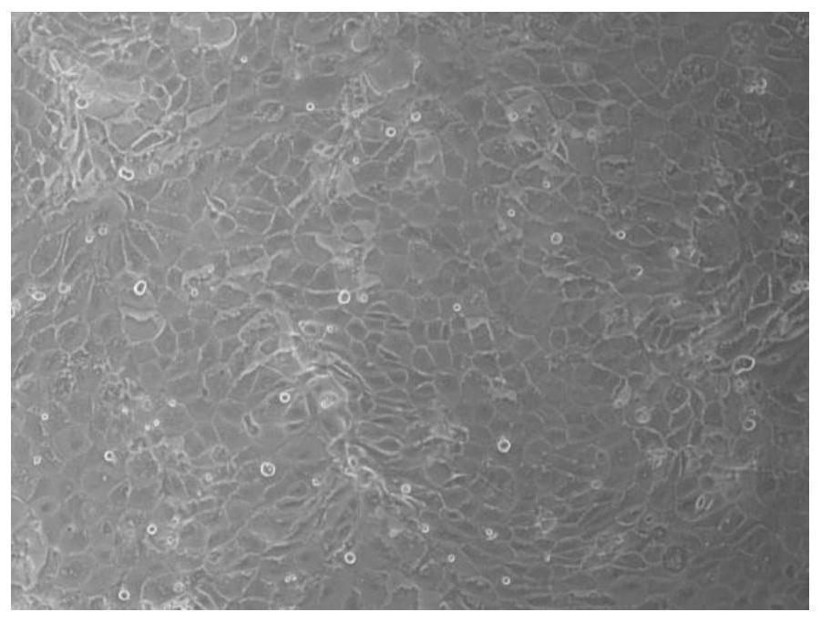
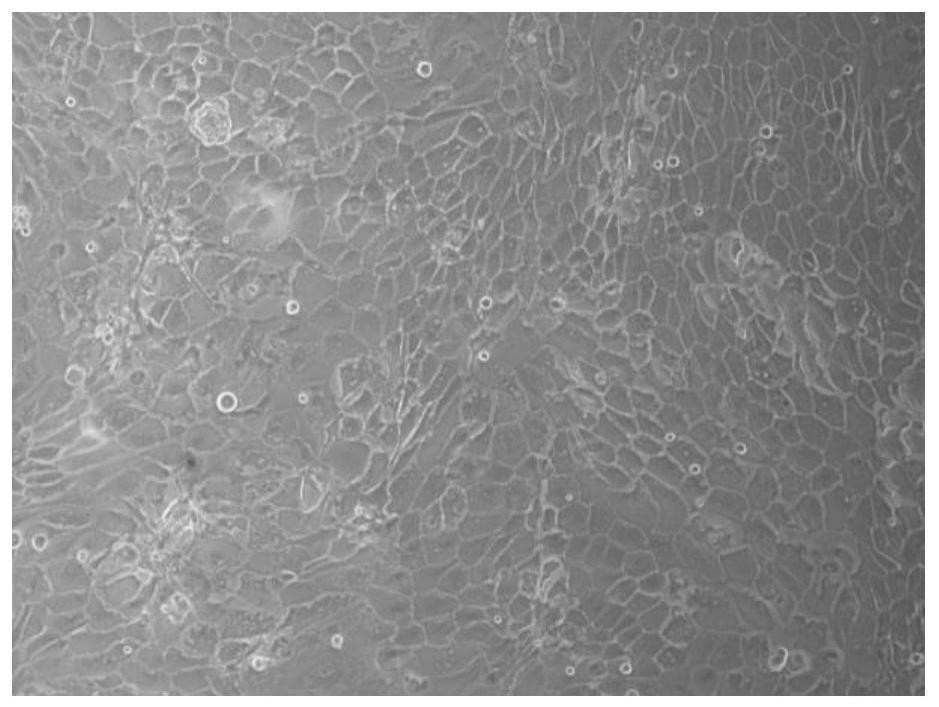
[0054] After the cells were transfected, the morphological changes were observed under a phase-contrast microscope, and compared with primary sheep endometrial epithelial cells (such as figure 1 )Compare.
[0055] The results showed that the transfected sheep endometrial epithelial cells ( figure 2 ) is similar to the primary cell morphology, the central nucleus of the cell is obvious, and the nucleolus is clearly visible. Most of the cells were polygonal or oval in shape, and the cells were arranged in a typical cobblestone-like arrangement, and there was contact inhibition between cells.
Embodiment 3
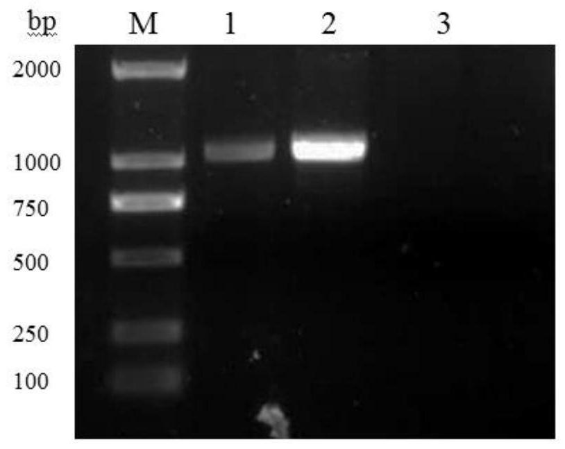
[0056] Example 3 RT-PCR detection of hTERT expression in transfected cells
[0057] (1) Design and synthesis of primers
[0058] Design a pair of specific detection primers according to the human telomerase reverse transcriptase gene reference sequence published in GenBank (accession number NM_001193376), the upstream and downstream primers are Forward: 5ˊ-AGAGGTCAGGCAGCATCGG-3ˊ, see SEQ ID NO.1; Reverse: 5ˊ-TGCGTTCTTGGCTTTCAGG-3ˊ, see SEQ ID NO.2, the size of the amplified fragment is 1156 bp, and the primers were synthesized by Shanghai Sangon Bioengineering Co., Ltd.
[0059] (2) Extraction of total cellular RNA
[0060] ① Inoculate the transfected cells and primary cells in a 6-well plate. After the cells are confluent, discard the culture medium and wash twice with PBS.
[0061] ②Add 750 μL Trizol reagent to each well, mix well by pipetting, and let stand at 4°C for 5 minutes.
[0062] ③Pipe the cell lysate into a 1.5 mL sterilized EP tube treated with DEPC water, cent...
PUM
 Abstract
Description
Claims
Application Information
 - R&D
- Intellectual Property
- Life Sciences
- Materials
- Tech Scout
- Unparalleled Data Quality
- Higher Quality Content
- 60% Fewer Hallucinations
Browse by: Latest US Patents, China's latest patents, Technical Efficacy Thesaurus, Application Domain, Technology Topic, Popular Technical Reports.
© 2025 PatSnap. All rights reserved.Legal|Privacy policy|Modern Slavery Act Transparency Statement|Sitemap|About US| Contact US: help@patsnap.com


