Staining method using fluorescence quenching time difference, automatic staining device, equipment and medium

A fluorescence staining and fluorescence quenching technology, applied in the field of cell staining, can solve the problems such as the inability of long-term preservation of images, the lack of various antibody analysis result maps, and the dependence of the ability to identify and distinguish.

Pending Publication Date: 2020-10-13
GUANGZHOU KINGMED DIAGNOSTICS CENT
View PDF8 Cites 2 Cited by
  • Summary
  • Abstract
  • Description
  • Claims
  • Application Information

AI Technical Summary

Problems solved by technology

[0003] Conventional HE staining has a wider range of observation than immunofluorescence staining, but the ability to identify and distinguish a specific cell or substance depends heavily on human experience and the limit ability of vision
The immunofluorescence staining method, using antibody technology, has the advantages of high sensitivity and high specificity to amplify the cells or substances to be detected, but the current immunofluorescence method has the disadvantage that the image cannot be stored for a long time, and when many staining antibodies are used, it is easy to The phenomenon of cross-interference between fluorescence spectra will affect the final result interpretation
However, the direct combination of immunofluorescence staining and HE staining will make the staining area unable to be completely consistent, which will lead to misjudgment of the staining observation area
Therefore, the existing staining methods can only observe 1-3 kinds of antibodies in HE staining images or immunofluorescence images, and lack the analysis results of comprehensive HE and multiple antibodies.

Method used


Image

  • Staining method using fluorescence quenching time difference, automatic staining device, equipment and medium
  • Staining method using fluorescence quenching time difference, automatic staining device, equipment and medium
  • Staining method using fluorescence quenching time difference, automatic staining device, equipment and medium

Examples


Embodiment Construction

[0061] The following will clearly and completely describe the technical solutions in the embodiments of the present invention with reference to the accompanying drawings in the embodiments of the present invention. Obviously, the described embodiments are only some, not all, embodiments of the present invention. Based on the embodiments of the present invention, all other embodiments obtained by persons of ordinary skill in the art without creative efforts fall within the protection scope of the present invention.

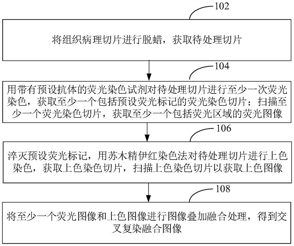
[0062] Such as figure 1 as shown, figure 1 It is a schematic flow chart of the staining method using the fluorescence quenching time difference in an embodiment, and the steps provided in this embodiment include:

[0063] Step 102, dewaxing the histopathological section to obtain the section to be processed.

[0064] Among them, the histopathological sections in this embodiment include tumor histopathological sections such as breast cancer histopathological secti...


PUM


Abstract

The invention discloses a staining method using fluorescence quenching time difference. The staining method comprises the following steps of: dewaxing a histopathological section to obtain a section to be treated, and carrying out fluorescence staining on the section to be treated at least once by using a fluorescence staining reagent with a preset antibody to obtain fluorescence staining sectionscomprising preset fluorescence labels; quenching the preset fluorescence labels generated in the previous fluorescence staining before carrying out the next fluorescence staining; and scanning each fluorescence staining section to obtain a fluorescence image; quenching the preset fluorescence labels, coloring and staining the section to be treated by using a hematoxylin eosin staining method to obtain a colored and stained section, and scanning the colored and stained section to obtain a colored image; and performing image superposition fusion processing on the at least one fluorescence imageand the colored image to obtain a cross-staining fusion image. Therefore, the staining method can meet requirements of the stained image on the observation range and the staining specificity. In addition, the invention further provides a staining device and staining equipment using the fluorescence quenching time difference, and a medium.

Description

technical field [0001] The invention relates to the technical field of cell staining, in particular to a staining method using the time difference of fluorescence quenching, an automatic staining device, equipment and a medium. Background technique [0002] Histopathological diagnosis is an important method in disease diagnosis. Histopathological diagnosis needs to be based on staining and observation of histopathological sections. Common staining methods include: HE (hematoxylin-eosin, hematoxylin-eosin) staining, immunohistochemical staining, immunofluorescence staining and other methods. [0003] Conventional HE staining has a wider range of observation than immunofluorescence staining, but the ability to identify and distinguish a specific cell or substance depends heavily on human experience and the limit ability of vision. The immunofluorescence staining method, using antibody technology, has the advantages of high sensitivity and high specificity to amplify the cells...

Claims


Application Information

Patent Timeline
Patent Type & Authority Applications(China)
IPC IPC(8): G01N1/31G01N21/64
CPCG01N1/31G01N21/6428G01N2021/6432
Inventor 车拴龙吴涛丁向东李映华丘伟松刘斯李东雄董西兰张胜群
Owner GUANGZHOU KINGMED DIAGNOSTICS CENT
Why Eureka
Try Eureka