Eureka AIR delivers breakthrough ideas for toughest innovation challenges, trusted by R&D personnel around the world.

Contrast agent for abdominal imageology examination

A contrast agent and abdominal technology, applied in the direction of X-ray contrast agent preparation, in vivo test preparations, radioactive carriers, etc., to achieve the effects of reducing absorption, improving safety, and easy application

Pending Publication Date: 2020-07-14
THE FIRST AFFILIATED HOSPITAL OF WENZHOU MEDICAL UNIV
View PDF3 Cites 0 Cited by
  • Summary
  • Abstract
  • Description
  • Claims
  • Application Information

AI Technical Summary

Problems solved by technology

[0013] In order to overcome the current bottleneck of lacking a contrast agent for abdominal imaging examination that can effectively suppress obesity, can be stored for a long time, is convenient to use, is safe and has no side effects, the purpose of the present invention is to provide a contrast agent for abdominal imaging examination

Method used


Image

  • Contrast agent for abdominal imageology examination
  • Contrast agent for abdominal imageology examination

Examples


Embodiment 1

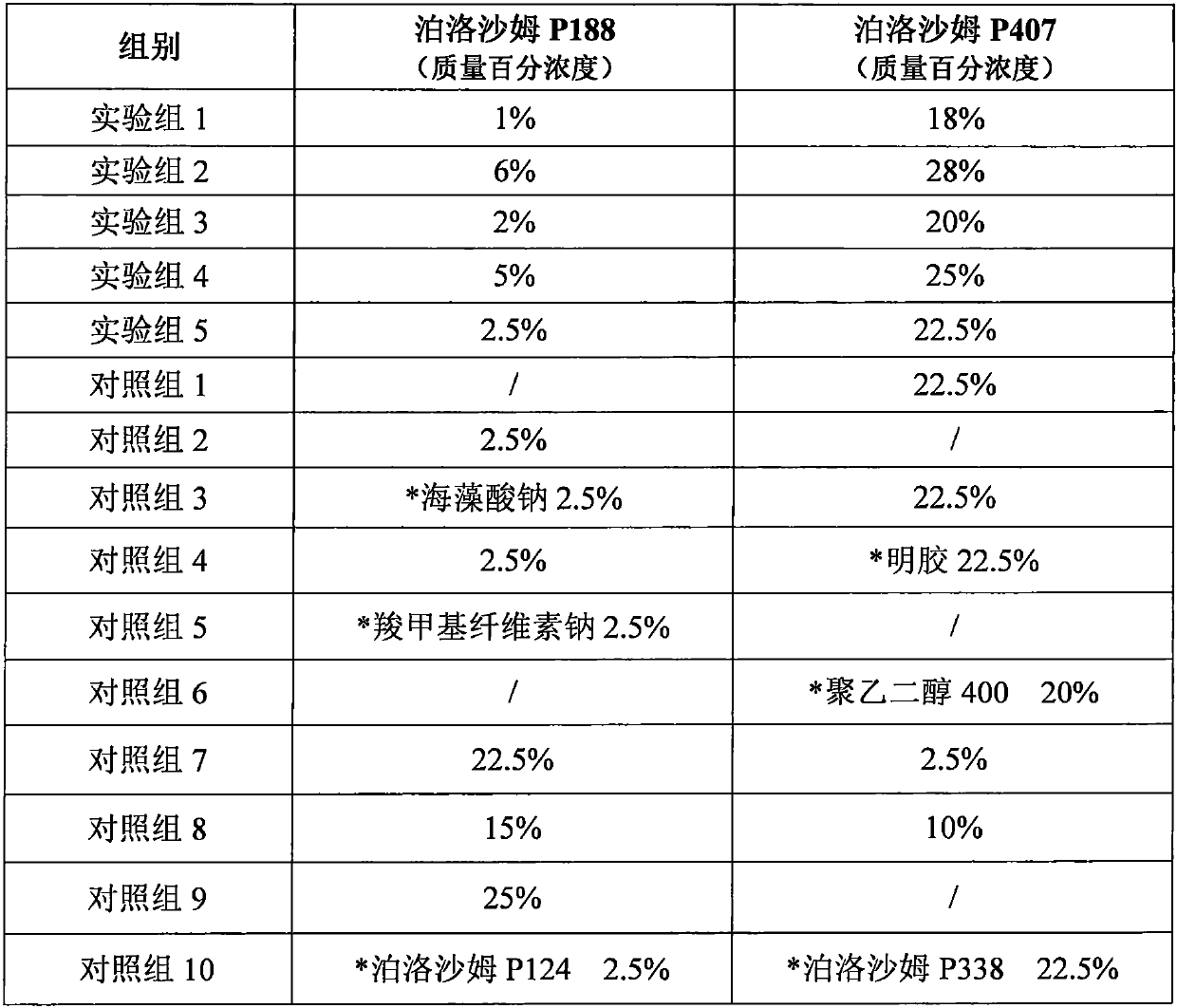
[0028] Embodiment 1 Preparation of contrast agent for abdominal imaging examination

[0029] Preparation of contrast agent for abdominal X-ray radiography in the experimental group: According to the composition of the experimental group in Table 1, each component was measured, and the contrast agent for abdominal X-ray radiography was prepared according to the following steps. Brief description of the preparation method: mix poloxamer P188 and poloxamer P407, add distilled water, mix well, place at 4°C for 12 hours, mix well again, add barium sulfate fine powder, disperse and mix well, and obtain abdominal X-ray radiography contrast agent.

[0030] Preparation of contrast medium for abdominal imaging examination in the control group: according to the composition of the control group in Table 1, the contrast medium for the abdominal imaging examination of the control group was prepared according to the method of the experimental group.

[0031] The composition of the abdominal...

Embodiment 2

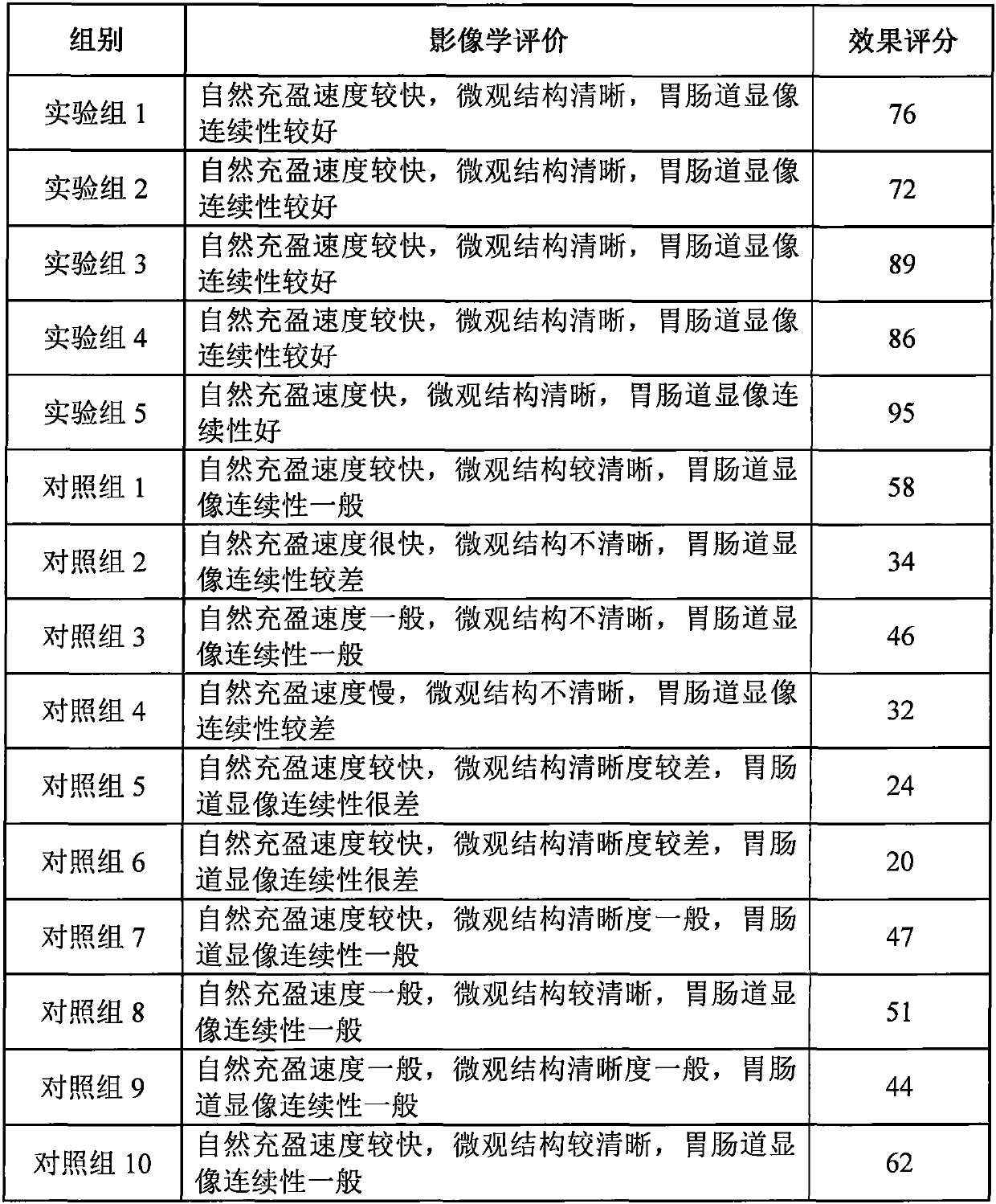
[0034] Embodiment 2 Evaluation of imaging effect of contrast medium in abdominal imaging examination

[0035] Gastric imaging: Beagle dogs (weight 3kg / dog) were fasted for 18-24 hours before imaging, food and debris in the stomach were removed, and the dog was anesthetized by intramuscular injection of Sumianxin. Before the contrast, the X-ray machine took a plain film, and then injected 30% weight / volume ratio of X-ray radiographic contrast agent through the gastric catheter, and poured it in at 10mL / kg body weight. Then turn the animal over carefully, so that the barium meal is fully coated on the stomach wall (turn over 3 to 5 times to the left and right, lift up and down 2 to 3 times). Then inject air into the stomach at a rate of 8-10 mL / kg body weight, take out or clamp the outer end of the gastric tube, and then turn it over 1-2 times left and right, and immediately adopt the ventral dorsal position (dorsal abdominal position) and left side (right side) filming.

[00...


PUM


Abstract

The invention provides a contrast agent for abdominal imageology examination. The contrast agent is a mixed solution which is composed of poloxamer P188 and poloxamer P407; and as an imaging contrastagent for stomach, intestinal tract, uterus and vagina and a matrix of the imaging contrast agent, the contrast agent is mixed with an imaging tracer agent and is used as the contrast agent for magnetic resonance imaging, positron emission computed tomography imaging, single photon emission computed tomography, computed tomography, combined imaging of positron emission computed tomography imagingand computed tomography and X-ray radiological imaging. The contrast agent for abdominal imageology examination disclosed by the invention has the characteristics that the contrast agent is simple andquick to use, the safety is high, the volume of the contrast agent keeps invariant, the adhesiveness is good, and natural filling and continuous filling of gastrointestinal tract can be realized.

Description

technical field [0001] The invention relates to an abdominal contrast agent, in particular to a contrast agent for abdominal imaging examination. Background technique [0002] Abdominal examination is the focus of medical imaging examinations. The traditional abdominal examination mainly focuses on gastrointestinal examination, while the uterus and vagina are the focus of gynecological abdominal examination. Currently commonly used examinations include: X-ray, computerized tomography (CT), magnetic resonance imaging (MRI), etc. In order to improve the resolution of images, it is necessary to cooperate with the application of contrast agents. [0003] In abdominal CT examination, gastrointestinal contrast medium is routinely used before the examination. At present, water contrast medium is advocated in upper abdominal CT examination, and positive contrast medium is required to fill the gastrointestinal tract in CT examination of the middle and lower abdomen and pelvis. Alth...

Claims


Application Information

Patent Timeline
IPC IPC(8): A61K49/04A61K49/12A61K51/06A61K101/02
CPCA61K49/04A61K49/0404A61K49/126A61K51/06
Inventor 赵应征鲁翠涛陈斌杨运俊徐荷林姚情
Owner THE FIRST AFFILIATED HOSPITAL OF WENZHOU MEDICAL UNIV
Why Eureka
Eureka Blog
Learn More