Positron tomoscan and reconstruction methods

A technology of positron tomography and scanning methods, which is applied in computerized tomography scanners, diagnosis, echo tomography, etc., and can solve problems such as low number of spatial response lines, low resolution of three-dimensional images, and bottleneck effects of PET equipment

Active Publication Date: 2019-12-13
FMI MEDICAL SYST CO LTD
View PDF8 Cites 3 Cited by
  • Summary
  • Abstract
  • Description
  • Claims
  • Application Information

AI Technical Summary

Problems solved by technology

As a result, in the actual scanning process, the number of effectively sampled spatial response lines in the axial direction is relatively low, and the actual resolution of the reconstructed 3D image in the axial direction is also lower than that in the tangential direction.
Therefore, when scanning relatively small fine objects, PET equipment will have a bottleneck effect in the axial resolution

Method used


Image

  • Positron tomoscan and reconstruction methods
  • Positron tomoscan and reconstruction methods
  • Positron tomoscan and reconstruction methods

Examples


Embodiment 1

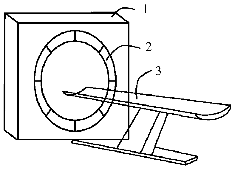
[0045] Such as Figure 1 to Figure 3 As shown, the positron emission tomography method of this embodiment has the following steps:

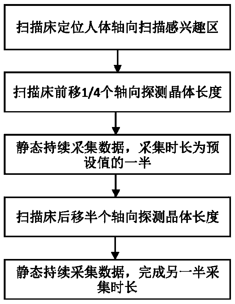
[0046] S101: During the PET scanning process, the scanning bed moves toward the PET device as a forward direction. The human body is moved by the scanning bed, and the PPET device is positioned to a predetermined axial region of interest, that is, the scanning bed is positioned to scan the region of interest in the axial direction of the human body;

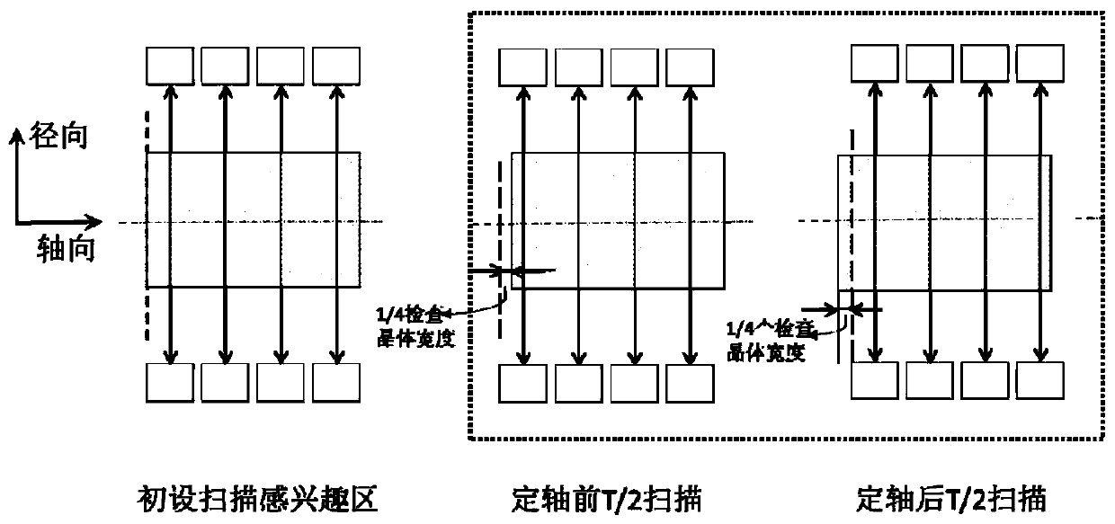
[0047] S102: moving the axial region of interest forward by 1 / 4 the crystal width of the axial detector;

[0048] S103: The PET device starts to collect data statically, and the continuous collection time is half of the preset single-bed scanning time;

[0049] S104: Move the region of interest backward by half the crystal width of the axial detector;

[0050] S105: Complete the static data collection task of the remaining single-bed scanning time.

[0051] The data output format of each static ...

Embodiment 2

[0067] Such as Figure 1 to Figure 3 As shown, the positron emission tomography method of this embodiment has the following steps:

[0068] S101: During the PET scanning process, the scanning bed moves toward the PET device as a forward direction. The human body is moved by the scanning bed, and the PPET device is positioned to a predetermined axial region of interest, that is, the scanning bed is positioned to scan the region of interest in the axial direction of the human body;

[0069] S102: moving the axial region of interest forward by 1 / 4 the crystal width of the axial detector;

[0070] S103: The PET device starts to collect data statically, and the continuous collection time is half of the preset single-bed scanning time;

[0071] S104: Move the region of interest backward by half the crystal width of the axial detector;

[0072] S105: Complete the static data collection task of the remaining single-bed scanning time.

[0073] The data output format of each static ...


PUM


Abstract

The invention provides a positron tomoscan method and a reconstruction method, and relates to the technical field of X-ray computed tomoscan imaging. The scanning method comprises the steps: a PET device is positioned to a predetermined axial region of interest; the axial region of interest is moved forward by 1 / 4 of the axial detector crystal width; the PET device starts to statically acquire data, and the continuous acquisition time is half of the preset single-bed scanning duration; the region of interest is moved backwards by half of the axial detector crystal width; and a static data acquisition task of remaining single-bed scanning time is completed. The reconstruction method comprises the step of combining two times of the scanning data on a coincidence event or on a sinogram. According to the scanning method, the positioning length of an original axial scanning region of interest is not changed; by controlling the small deviation of the scanning bed, two times of sub-scanning are carried out in the region of interest, the single scanning time is half of the original scanning time, and more fined spatial distribution information in the axial direction can be obtained, so that the axial imaging resolution rate of the PET system is improved.

Description

technical field [0001] The present invention relates to X-ray computed tomography imaging (PET / CT, PET / MRI), in particular to a positron emission tomography and reconstruction method. Background technique [0002] PET (Positron Emission Tomography) equipment is an advanced functional imaging tool in nuclear medicine imaging. The nuclide decays in the organism and emits a positron, which annihilates with the electrons of nearby substances to produce a pair of photons with equal energy and opposite direction; the detector of the PET device captures this pair of high-energy photons, which is called a coincidence event , form a line of response (LOR) in the scanning space, convert the space and time signals of the pair of photons into electronic information, and store them in the storage medium. After a period of cumulative collection and enough coincident events are obtained, a reconstruction algorithm is used to reconstruct the spatial distribution of radioactive substances i...

Claims


Application Information

Patent Timeline
IPC IPC(8): A61B6/03A61B6/00
CPCA61B6/037A61B6/5211
Inventor 褚政王瑶法高思泽叶宏伟
Owner FMI MEDICAL SYST CO LTD
Why Eureka
Try Eureka