SERS-fluorescent dual mode probe based on DNA chain replacement, and preparation method and application of SERS-fluorescent dual mode probe
A DNA chain and fluorescence technology, applied in the field of bioanalytical chemistry, can solve the problems of unstable signal, inability to accurately observe the position and change of active substances in cells, complicated operation, etc., and achieve the effect of stable nature.
- Summary
- Abstract
- Description
- Claims
- Application Information
AI Technical Summary
Problems solved by technology
Method used
Image
Examples
Embodiment 1
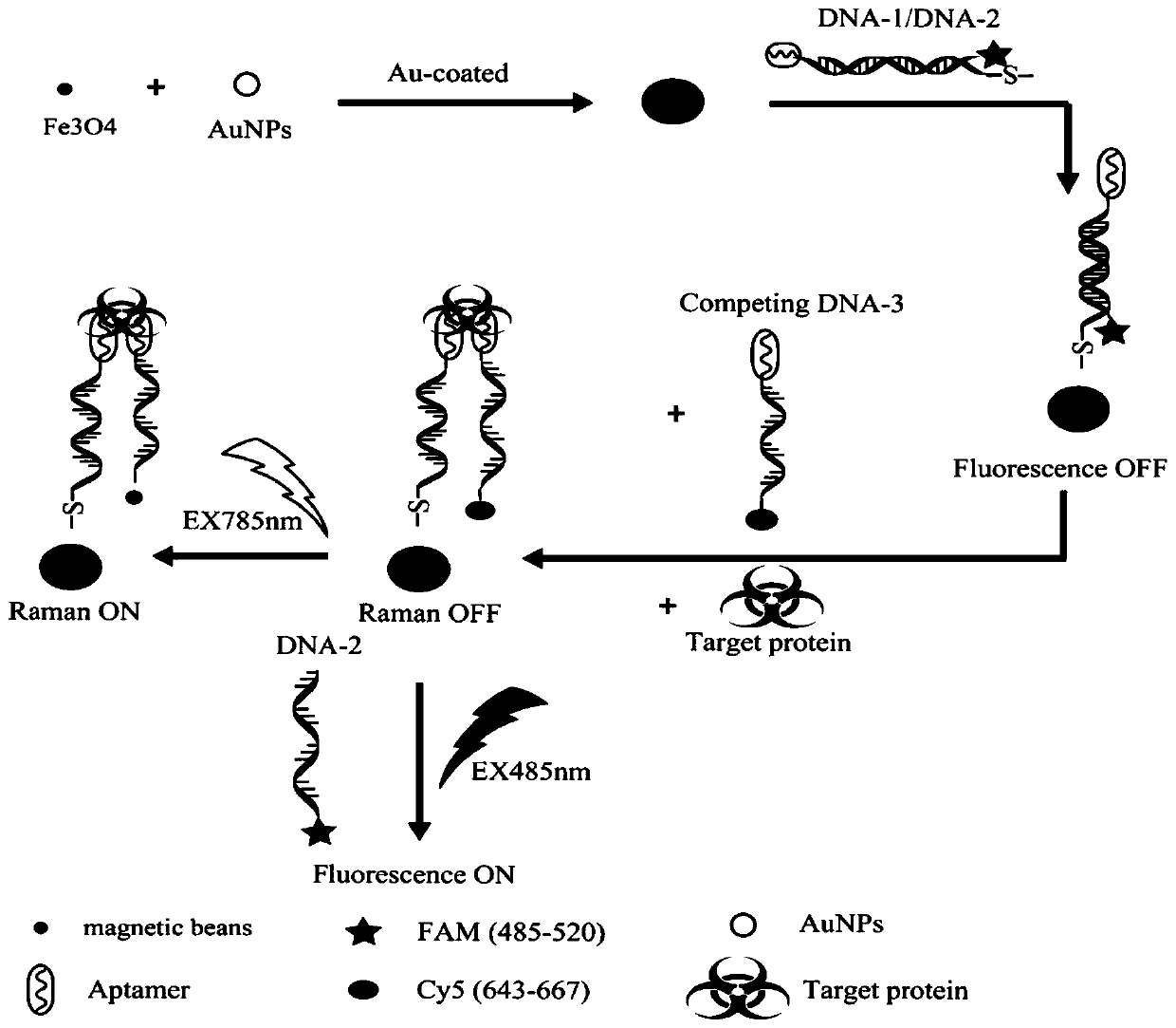
[0035] A method for preparing a SERS-fluorescent dual-mode probe based on DNA strand displacement, such as figure 1 As shown, it specifically includes the following steps:
[0036] 1) Fe 3 o 4 Preparation: Take 1~5g FeCl 2 4H 2 O and 1~5g FeCl 3 ·6H 2 Dissolve O in 20mL ultrapure water, filter through filter paper and dilute 10-30mL, and after a few minutes of nitrogen gas, add 10-30mL ammonia water to the above solution drop by drop, in N 2 React under protection for 30 minutes and age for 1 to 3 hours. After the reaction, the heat source was removed and cooled to room temperature, separated by a magnet, washed repeatedly with water and ethanol several times, dried in an oven overnight and ground into powder for later use.
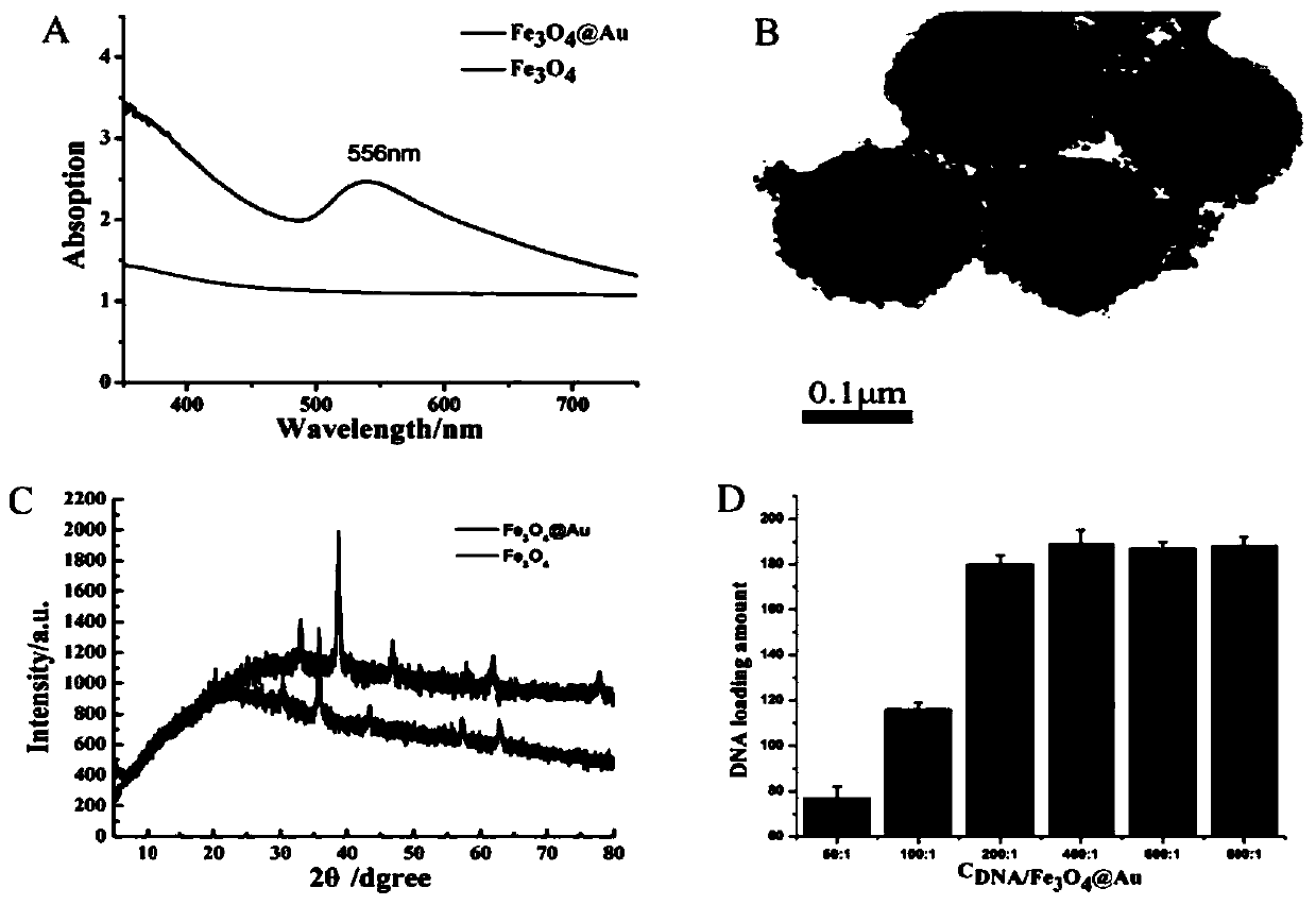
[0037] 2) Fe 3 o 4 @Au's preparation:
[0038] 2a) Fe 3 o 4 - Preparation of DA: weigh 10-30mg Fe 3 o 4 Dissolve in THF, add 2mL dopamine hydrochloride aqueous solution, sonicate evenly, stir overnight at room temperature, centrifuge to remo...
Embodiment 2
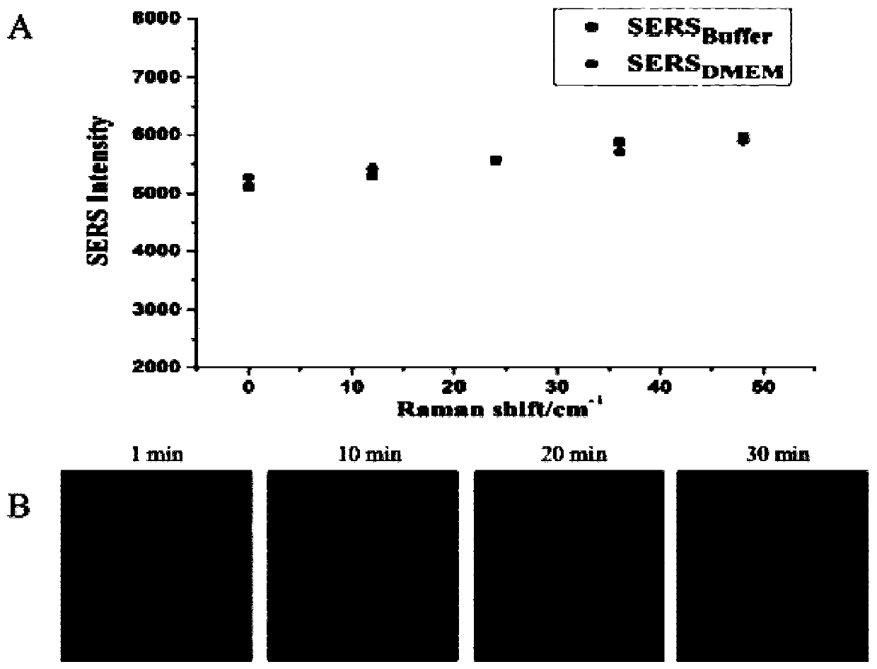
[0050] SERS-fluorescent dual-mode probes for SERS quantitative detection of VEGF in vitro, such as Figure 4 shown.
[0051] The Raman detection in the constructed detection method includes the following steps: In this embodiment, human melanoma cell A375 is taken as an example. After the A375 cells grow to the logarithmic phase, the cells are divided into 6 Inoculate 100 μL onto a sterile quartz plate at a density of 1 / mL, incubate for 24 hours until the wall is completely adhered, suck out the old medium, add DNA containing DNA and mix well 1-2 Modified Fe 3 o 4 @Au probe medium 1mL, co-culture with cells for 24h, suck out the old medium and wash it twice with PBS, then add DNA mixed with the same concentration 3 The basal medium was co-cultured for 0, 0.5, 1, 1.5, 2, 2.5 and 3 hours respectively, then the old medium was aspirated, washed twice with PBS, and SERS imaging was performed immediately.
[0052] in, Figure 4 -A is the SERS signal diagram of the SERS-fluoresc...
Embodiment 3
[0054] Qualitative fluorescence imaging analysis of SERS-fluorescent dual-mode probes, such as Figure 5 shown.
[0055] Fluorescence detection in the constructed detection method includes the following steps: In this embodiment, human melanoma cell A375 is taken as an example. After the cells grow to the logarithmic growth phase, the cells are 6 Inoculate 100 μL onto a fluorescent confocal dish at a density of cells / mL, incubate for 24 hours and wait for complete attachment, suck out the old medium, add DNA containing DNA and mix well 1-2 Modified Fe 3 o 4 @Au medium 1mL, co-culture with cells for 24h, suck out the old medium and wash it twice with PBS, then add DNA mixed with the same concentration 3 The basal medium was co-cultured for 0, 1, 10, 20, and 30 min respectively; then the old medium was aspirated, washed twice with PBS, and immediately performed fluorescence imaging.
[0056] The results showed that due to DNA 3 Displacement of DNA by strand displacement and...
PUM
| Property | Measurement | Unit |
|---|---|---|
| particle diameter | aaaaa | aaaaa |
| particle diameter | aaaaa | aaaaa |
Abstract
Description
Claims
Application Information
 - R&D
- Intellectual Property
- Life Sciences
- Materials
- Tech Scout
- Unparalleled Data Quality
- Higher Quality Content
- 60% Fewer Hallucinations
Browse by: Latest US Patents, China's latest patents, Technical Efficacy Thesaurus, Application Domain, Technology Topic, Popular Technical Reports.
© 2025 PatSnap. All rights reserved.Legal|Privacy policy|Modern Slavery Act Transparency Statement|Sitemap|About US| Contact US: help@patsnap.com


