
A dyeing method for observing the internal microstructure of plants

A technology of microstructure and staining method, which is applied in testing plant materials, preparation of test samples, instruments, etc., can solve the problems of large workload of freehand sectioning, easy evaporation of sectioning, long time, etc., and achieve flexible dyeing experiment time. , reduce workload, slice complete effect

Active Publication Date: 2021-12-28
YANGTZE UNIVERSITY
View PDF1 Cites 0 Cited by
  • Summary
  • Abstract
  • Description
  • Claims
  • Application Information

AI Technical Summary

Problems solved by technology

But there are two prominent problems in the prior art. One is that the fresh plant material is put into distilled water after being sliced ​​by hand, and it needs to be observed immediately. In actual operation, the time for slicing by hand is too long, and the distilled water is easy to evaporate and make the slice shrinkage; the second is to observe the anatomical structures such as aerenchyma, lignification, suberinization, and Caspian's band. A total of 4 intact sections are required, and 4 times of staining are required. The workload of freehand sectioning is relatively large

Method used


Image

  • A dyeing method for observing the internal microstructure of plants

Examples


Embodiment 1

[0021] The invention provides a dyeing method for observing the internal microstructure of plants, comprising the steps of:

[0022] Step 1. Making freehand slices: use a blade to cut the small piece of plant root into slices with a thickness of 0.2-0.5 mm.

[0023] Step 2. Put the freehand slices into glycerin and save them for later use;

[0024] Step 3, drug preparation:

[0025] (1) Preparation of 0.05% (w / v) toluidine blue (TBO) dye solution: Take 0.05g TBO, dissolve in 100mL distilled water, mix well to prepare 0.05% TBO.

[0026] (2) Preparation of 5% (w / v) phloroglucinol solution: take 4 to 5 g of phloroglucinol, dissolve it in 100 ml of 95% alcohol, mix well to form phloroglucinol-alcohol solution, and prepare it now Use (solution yellow-brown failure).

[0027] (3) Preparation of 0.1% (w / v) Sudan 7B solution: Weigh 50 mg of Sudan 7B and dissolve it in 25 mL of PEG-300, then cool in a 90°C water bath for 1 hour, add an equal volume of 90% glycerol, and store at roo...

experiment example

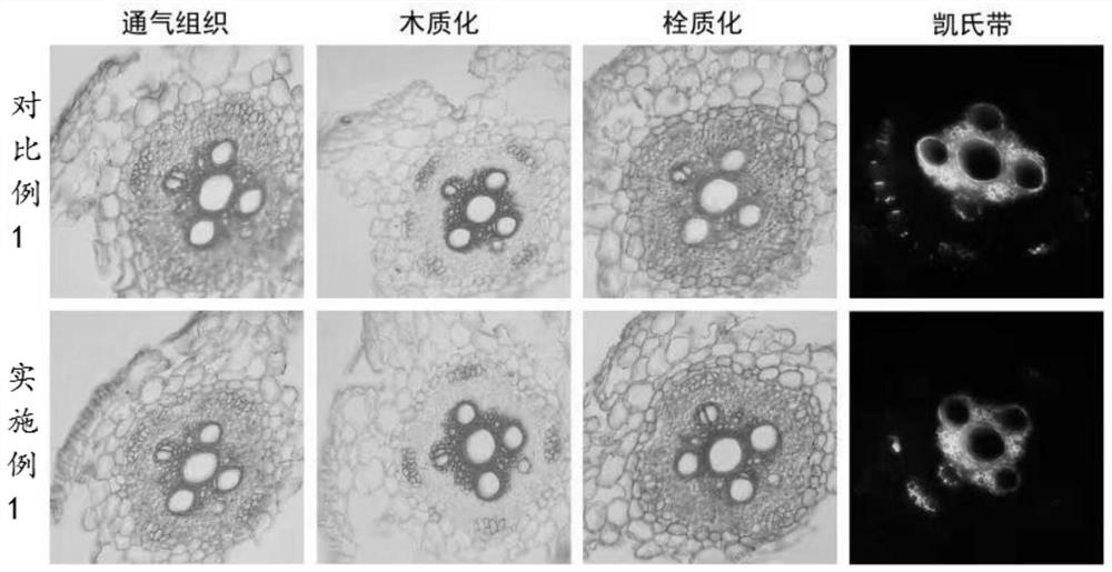
[0040] Record and count the microstructural diagrams of the aerenchyma, lignification, suberinization, and Caspian bands obtained in Example 1 and Comparative Example 1. figure 1 shown.

[0041] Depend on figure 1 It can be seen that both Example 1 and Comparative Example 1 can clearly see the microstructure of aerenchyma, lignification, suberinization, and Kjeldahl bands.

[0042] The method of the embodiment 1 of the present invention has obtained the same or similar results through repeated trials and experiments, showing that the method is stable and reliable, the observation result is clear, and the internal structure of the root can be seen clearly. Compared with the existing method, it can be carried out successively. 4 staining observations (air tissue, lignification, suberinization and Kjeldahl band), greatly reducing the workload of freehand sectioning, saving experimental materials and improving experimental efficiency.


PUM


Abstract

The invention provides a dyeing method for observing the internal microstructure of plants, comprising: (1) slicing by hand; (2) putting the slicing by hand into glycerin and storing it for later use; (3) placing the slicing by hand on a glass slide, and adding After staining the freehand sections with toluidine blue, add water dropwise and observe the aerenchyma under a microscope; (4) add phloroglucinol for staining, then add concentrated hydrochloric acid for staining, and observe lignification under a microscope; After washing with 75% ethanol, add Sudan 7B dropwise for staining, then add water dropwise, and observe suberization under a microscope; Water was added dropwise, and the Caspian bands were observed under a fluorescence microscope. This method can store freehand slices in glycerin and use them anytime, which makes the experiment time more flexible, and only one intact slice can be stained and observed four times successively, which reduces the workload of freehand slices, saves experimental materials, and improves the experimental efficiency.

Description

technical field [0001] The invention relates to the technical field of plant microstructure, in particular to a dyeing method for observing the internal microstructure of plants. Background technique [0002] Freehand sectioning of fresh plant tissue allows the microstructure of the plant interior to be observed with an optical microscope. The microstructure of the plant interior usually includes aerenchyma, lignification, suberinization, and Caspian bands. The study of anatomy is very important for plant classification, development, ecology, physiology, etc. But there are two prominent problems in the prior art. One is that the fresh plant material is put into distilled water after being sliced ​​by hand, and it needs to be observed immediately. In actual operation, the time for slicing by hand is too long, and the distilled water is easy to evaporate and make the slice Shrinkage; the second is to observe the anatomical structures such as aerenchyma, lignification, suberi...

Claims


Application Information

Patent Timeline
Patent Type & Authority Patents(China)
IPC IPC(8): G01N1/30G01N21/84
CPCG01N1/30G01N21/84G01N2021/8466
Inventor 朱进彭玉全李文静
Owner YANGTZE UNIVERSITY
Learn More