Adriamycin-carrying lipid nanoscale ultrasound contrast agent targeting tumor-related fibroblasts and preparation method of adriamycin-carrying lipid nanoscale ultrasound contrast agent
A technology of fibroblasts and ultrasound contrast agents, applied in echo/ultrasound imaging agents, medical preparations containing active ingredients, anti-tumor drugs, etc., to achieve high biological safety, excellent imaging capabilities, and enhanced imaging capabilities Effect
- Summary
- Abstract
- Description
- Claims
- Application Information
AI Technical Summary
Problems solved by technology
Method used
Image
Examples
Embodiment 1
[0058] Example 1: Synthesis and identification of targeting material DSPE-PEG-FH
[0059] Synthesis of targeting material DSPE-PEG-FH:
[0060] The targeting material DSPE-PEG-FH was prepared by condensation reaction of amino group and carboxyl group. Mix DSPE-PEG-NHS, FH short peptide, and HBTU with anhydrous DMF according to a certain ratio (molar ratio 3:1:3), and add triethylamine to adjust the pH to 8-9. Gentle stirring reaction at room temperature for 2h. After mixing water, 1,2-ethanedithiol, triisopropylsilane, and trifluoroacetic acid (1:2:2:95, v / v / v / v), add the above reaction according to the volume ratio of 1:5 solution, used to remove the protective group of the short peptide, separated DSPE-PEG-FH with ether, and dialyzed the separated product for 48 hours, with a molecular weight cut-off value of 3500Da. Finally, the final product DSPE-PEG-FH was lyophilized and stored at -20°C.
[0061] Identification of targeting material DSPE-PEG-FH:
[0062] The final p...
Embodiment 2
[0063] Example 2: Preparation of lipid nanoscale ultrasound contrast agent targeting CAF and carrying DOX
[0064] (1) Weigh 0.1ml of propylene glycol and 0.1ml of glycerin and place them in a 1.5ml EP tube, add 5mgDPPC, 2mg of DSPE-PEG-NH2, 0.5mg of DSPE-PEG-FITC, and 0.5mg of DSPE-PEG-FH prepared in Example 1 , PBS to a volume of 1ml; the PBS is a phosphate buffer saline, and its components are: Na 2 HPO 4 8mM, KH 2 PO 4 2mM, pH 7.2;
[0065] (2) Pass perfluoropropane into the mixture in step (1) to replace the air, place the centrifuge tube in a constant temperature water bath at 60°C and heat until the lipid is completely dissolved;
[0066] (3) Add DOX (0.5, 1.0, 1.5, 2.0 mg / ml) to the mixture in step (2), shake it mechanically for 90 seconds, take it out, centrifuge at 300 rpm for 5 minutes, and take out the lower layer of liquid, which is the prepared nano-scale ultrasound contrast agent.
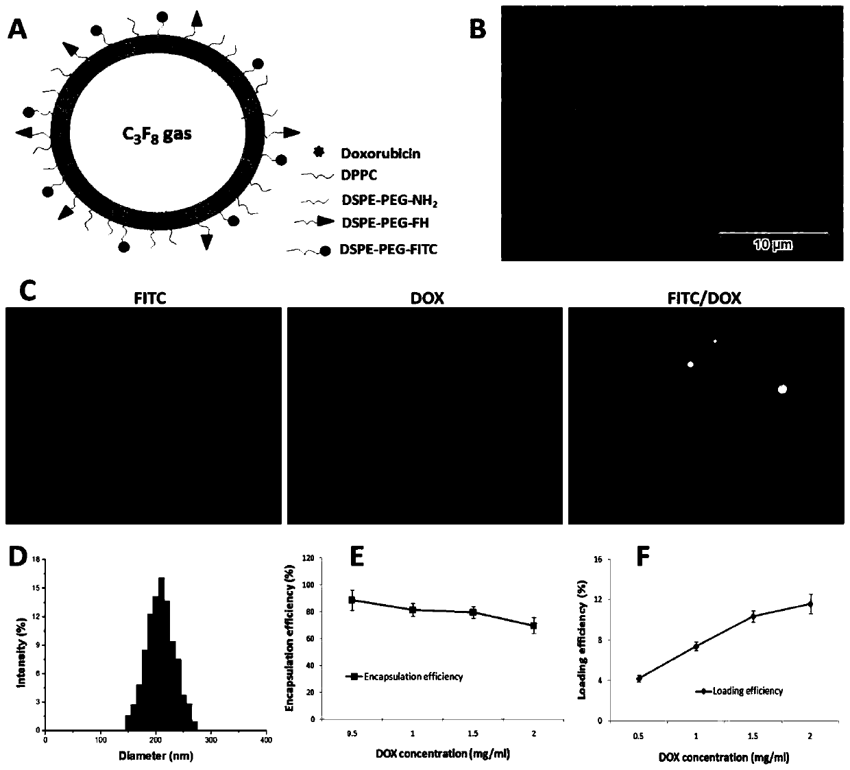
[0067] figure 2 A shows the schematic diagram of nano-scale ultrasound ...
Embodiment 3
[0071] Example 3: Experiment for Determination of In Vitro Developing Ability of Nanoscale Ultrasound Contrast Agent
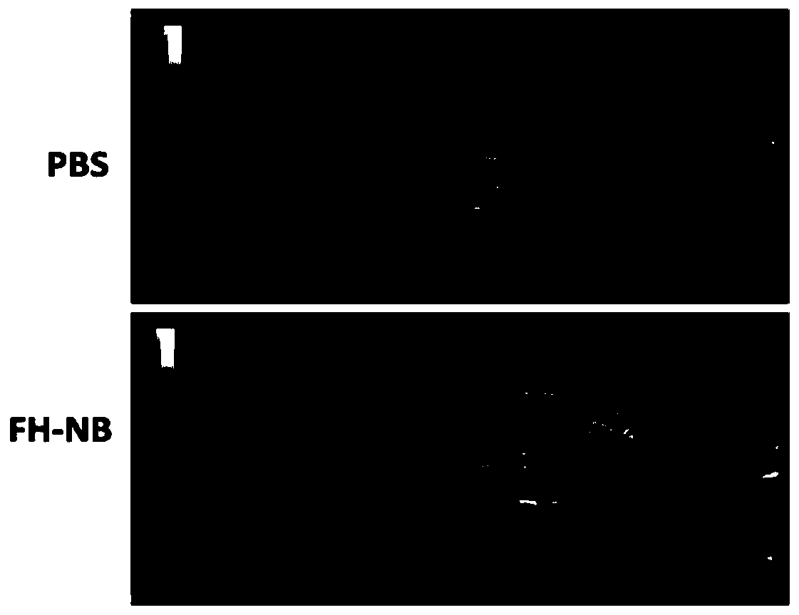
[0072] The nano-scale ultrasound contrast agent prepared in Example 2 was placed in the round hole made of agar gel, and the contrast mode of the GE logiqE9 ultrasonic diagnostic instrument was used, the frequency was 9.0MHz, the mechanical index (MI) was 0.12, and the two-dimensional and ultrasound contrast modes were used. Simultaneously observe, adjust parameter settings, store image data with the internal workstation of the ultrasonic instrument, and use PBS as the control group. image 3 The results showed that enhanced imaging appeared in the nanoscale ultrasound contrast agent group, but no enhanced imaging occurred in the control PBS group.
PUM
 Abstract
Description
Claims
Application Information
 - R&D Engineer
- R&D Manager
- IP Professional
- Industry Leading Data Capabilities
- Powerful AI technology
- Patent DNA Extraction
Browse by: Latest US Patents, China's latest patents, Technical Efficacy Thesaurus, Application Domain, Technology Topic, Popular Technical Reports.
© 2024 PatSnap. All rights reserved.Legal|Privacy policy|Modern Slavery Act Transparency Statement|Sitemap|About US| Contact US: help@patsnap.com


