Fluorescence immunoassay method based on carbon quantum dots
A technology of carbon quantum dots and fluorescence, applied in the field of fluorescence immunoassay based on carbon quantum dots, can solve the problems of undeveloped application potential and few carbon dots
- Summary
- Abstract
- Description
- Claims
- Application Information
AI Technical Summary
Problems solved by technology
Method used
Image
Examples
Embodiment 1
[0072] The synthesis of embodiment 1 fluorescent carbon quantum dots
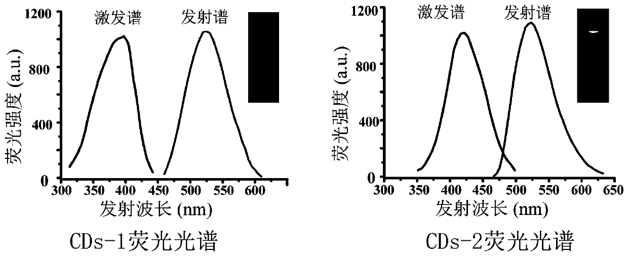
[0073] 5 g of citric acid and 7 g of thiourea were added to 15 mL of water. Stir continuously to dissolve the compound, place the mixed solution in a microwave reactor, and heat it at 800W for 7 minutes until the solution changes from colorless to dark brown solid, indicating that the reactant is carbonized and carbon quantum dots are formed. After cooling to room temperature, 25 mL of water was added to dissolve the product. The prepared carbon quantum dots were filtered through a 0.22 μm membrane to remove large particles, and dialyzed in ultrapure water for at least 24 h using a dialysis membrane (3500 Da) to remove unreacted small molecules. The prepared carbon quantum dot solution was separated and purified by Sephadex G25 column chromatography, and the collected carbon quantum dot solution with a maximum excitation wavelength of 370 nm and a maximum emission wavelength of 520 nm was named CDs-1.
[...
Embodiment 2
[0076] Embodiment 2 Amantadine fluorescent immunoassay detection
[0077] 1) Dilute the amantadine artificial antigen with coating solution to a concentration of 3.3×10 –5 mg / mL, add 100 μL / well into a 96-well ELISA plate, incubate at 4°C for 12 hours, then wash the plate with washing solution;
[0078] 2) Add blocking solution at 150 μL / well for blocking, incubate at 37°C for 1 hour, and wash the plate with washing solution;
[0079] 3) Add specific anti-amantadine antibody (concentration: 2.5×10 –5 mg / mL) and 50 μL / well of the sample solution to be tested, incubated at 37°C for 0.5 h, the antigen-antibody specific binding reaction occurs, and then the plate is washed with washing solution;
[0080] 4) Add horseradish peroxidase or alkaline phosphatase-labeled secondary antibody (concentration: 0.1 mg / mL) at 100 μL / well, incubate at 37 °C for 0.5 h to form enzyme-labeled immune complexes, and wash the plate with washing solution;
[0081] 5) Add horseradish peroxidase substr...
Embodiment 3
[0086] Embodiment 3 Aflatoxin B1 fluorescence immunoassay detection
[0087] 1) Dilute the artificial antigen of aflatoxin B1 with the coating solution to a concentration of 2.3×10 –5 mg / mL, add 100 μL / well into a 96-well ELISA plate, incubate at 4°C for 12 hours, then wash the plate with washing solution;
[0088] 2) Add blocking solution at 150 μL / well for blocking, incubate at 37°C for 1 hour, and wash the plate with washing solution;
[0089] 3) Add specific anti-aflatoxin B1 antibody (concentration: 2.1×10 –5 mg / mL) and 50 μL / well of the sample solution to be tested, incubated at 37°C for 0.5 h, the antigen-antibody specific binding reaction occurs, and then the plate is washed with washing solution;
[0090] 4) Add horseradish peroxidase or alkaline phosphatase-labeled secondary antibody (concentration: 0.1 mg / mL) at 100 μL / well, incubate at 37 °C for 0.5 h to form enzyme-labeled immune complexes, and wash the plate with washing solution;
[0091] 5) Add horseradish p...
PUM
 Abstract
Description
Claims
Application Information
 - Generate Ideas
- Intellectual Property
- Life Sciences
- Materials
- Tech Scout
- Unparalleled Data Quality
- Higher Quality Content
- 60% Fewer Hallucinations
Browse by: Latest US Patents, China's latest patents, Technical Efficacy Thesaurus, Application Domain, Technology Topic, Popular Technical Reports.
© 2025 PatSnap. All rights reserved.Legal|Privacy policy|Modern Slavery Act Transparency Statement|Sitemap|About US| Contact US: help@patsnap.com


