
Colorimetric kit for rapidly detecting chicken egg-yolk antibody

A technology of egg yolk antibody and polymer, applied in the field of biological detection, can solve the problems of low detection rate of antibodies, great influence of antibody interference, and long detection time.

Active Publication Date: 2019-03-19
JILIN UNIV
View PDF1 Cites 2 Cited by
  • Summary
  • Abstract
  • Description
  • Claims
  • Application Information

AI Technical Summary

Problems solved by technology

[0005] The purpose of the present invention is to solve the problems of traditional antibody detection methods such as long detection time, great influence by antibody interference, and low antibody detection rate, and provide an efficient, sensitive, fast, simple, high specificity and economical method for detecting chicken egg yolk antibodies Colorimetric Kit

Method used


Image

  • Colorimetric kit for rapidly detecting chicken egg-yolk antibody
  • Colorimetric kit for rapidly detecting chicken egg-yolk antibody
  • Colorimetric kit for rapidly detecting chicken egg-yolk antibody

Examples


Embodiment 1

[0027] Example 1 Magnetic Nanoparticles Fe 3 o 4 Preparation of NPs

[0028] With vigorous stirring, 2.7 g FeCl 3 ·6H 2 O and 7.2 g NaAc were dissolved in 100 mL of ethylene glycol to obtain a homogeneous brown-yellow solution; the homogeneous brown-yellow solution was transferred to a Teflon-lined reactor and sealed, and reacted in an oven at 200 °C for 8 h , after the reaction kettle was cooled to room temperature, black magnetite particles were separated from the reaction solution by means of permanent magnets; the black magnetite particles were washed alternately with absolute ethanol and ultrapure water for 6 to 7 times, and vacuumed at 60 °C After drying for 12 h, the magnetic nanoparticles Fe 3 o 4 NPs;

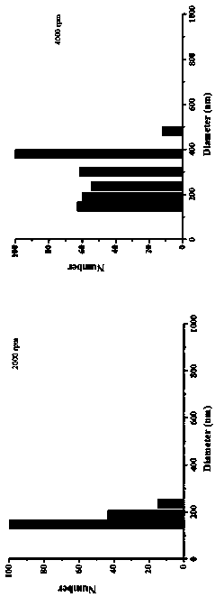
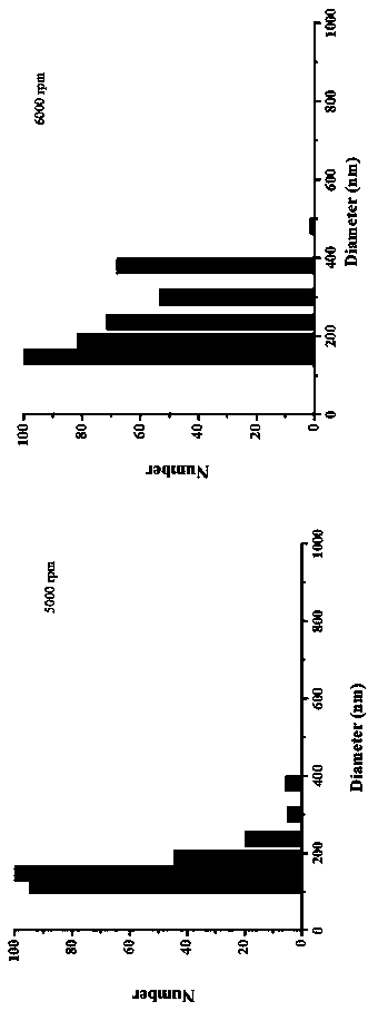
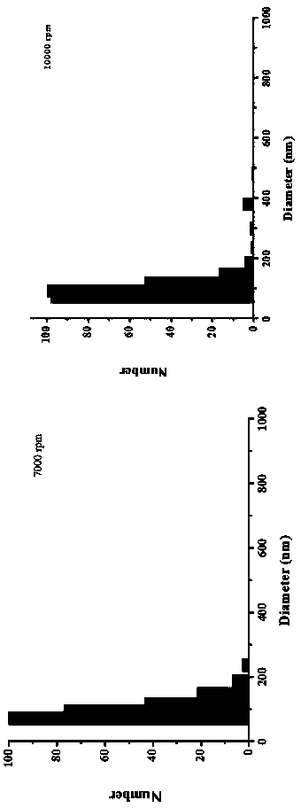
[0029] In order to obtain magnetic nanoparticles with uniform size, this study adopts the centrifugal precipitation method to separate the magnetic nanoparticles, and uses dynamic light scattering (DLS) to characterize the obtained magnetic nanoparticles. The r...

Embodiment 2

[0030] Example 2 Magnetic nanoparticles Fe 3 o 4 Preparation of NPs

[0031] With vigorous stirring, 2.7 g FeCl 3 ·6H 2 O and 7.2 g NaAc were dissolved in 100 mL of ethylene glycol to obtain a homogeneous brown-yellow solution; the homogeneous brown-yellow solution was transferred to a Teflon-lined reactor and sealed, and reacted in an oven at 180 °C for 10 h , after the reaction kettle was cooled to room temperature, black magnetite particles were separated from the reaction solution by means of permanent magnets; the black magnetite particles were washed alternately with absolute ethanol and ultrapure water for 6 to 7 times, and vacuumed at 60 °C After drying for 18 h, the magnetic nanoparticles Fe 3 o 4 NPs.

Embodiment 3

[0032] Example 3 Magnetic nanoparticles Fe 3 o 4 Preparation of NPs

[0033] With vigorous stirring, 2.7 g FeCl 3 ·6H 2 O and 7.2 g NaAc were dissolved in 100 mL of ethylene glycol to obtain a homogeneous brown-yellow solution; the homogeneous brown-yellow solution was transferred to a Teflon-lined reactor and sealed, and reacted in an oven at 200 ° C for 18 h. After the reactor is cooled to room temperature, black magnetite particles are separated from the reaction solution by means of a permanent magnet; the black magnetite particles are alternately washed several times with absolute ethanol and ultrapure water, and vacuum-dried at 55°C for 15 hours. Magnetic nanoparticles Fe 3 o 4 NPs.


PUM


Abstract

The invention discloses a colorimetric kit for rapidly detecting a chicken egg-yolk antibody. The colorimetric kit comprises a magnetic molecularly imprinted polymer. The magnetic molecularly imprinted polymer is prepared by the following method steps: 1) preparing Fe3O4 NPs with uniform size; 2) adding the Fe3O4 NPs into Tris buffer, performing ultrasonic dispersion, adding 1-5mg of egg yolk antibody and 2-5mg of dopamine, and mechanically stirring at a room temperature for 6-12 hours; 3) washing, and performing vacuum drying, thereby obtaining the magnetic molecularly imprinted polymer. Themagnetic nano-particle Fe3O4 NPs is prepared by the following steps: dissolving 2.7g of FeCl3*6H2O and 7.2g of NaAc into 100mL of ethylene glycol while intense stirring, sealing, reacting at a high temperature, cooling to a room temperature, and performing magnetic separation so as to obtain black magnetite particles; and washing, and performing vacuum drying, thereby obtaining the magnetic nano-particle Fe3O4 NPs.

Description

technical field [0001] The invention belongs to the field of biological detection, in particular to a colorimetric kit for rapidly detecting chicken yolk antibody. Background technique [0002] Yolk antibody (immunoglobulin of egg yolk, IgY), also known as egg yolk immunoglobulin, refers to the use of specific antigens to stimulate poultry B lymphocytes, which differentiate into plasma cells and secrete specific antibodies into the blood circulation. In the ovary, it gradually accumulates in the egg cells to form specific immunoglobulins, namely yolk antibodies. Yolk antibody IgY was initially thought to be similar in structure to mammalian immunoglobulin IgG. With the in-depth study of IgY, it was found that there were some differences in structure and properties between the two. The basic structure of IgY is similar to that of IgG, both of which are composed of two heavy chains (H chains) and two light chains (L chains). The differences are: ①IgY heavy chain consists of ...

Claims


Application Information

Patent Timeline
IPC IPC(8): G01N21/78G01N21/33
CPCG01N21/33G01N21/78
Inventor 王娟郭潇潇刘玉申郭媛媛姚硕魏源韩颖赵超李娟
Owner JILIN UNIV
Learn More