Method for constructing engineered artery blood vessel in vivo by taking melt-spun fibers as framework

A melt spinning, arterial blood vessel technology, applied in the field of medical devices, can solve the problems of easy formation of acute thrombus, body damage, vascular stenosis, etc., and achieve the effects of avoiding aneurysm formation, good biocompatibility, and reducing restenosis rate.

Inactive Publication Date: 2018-09-14
NANKAI UNIV
View PDF6 Cites 1 Cited by
  • Summary
  • Abstract
  • Description
  • Claims
  • Application Information

AI Technical Summary

Problems solved by technology

However, the use of autologous blood vessels requires a second operation, which not only causes damage to the body and additional costs, but also in some cases the patient cannot provide autologous blood vessels
Commercial artificial blood vessels (polyester and expanded polytetrafluoroethylene) show good patency in large-diameter vascular grafts, but small-diameter blood vessels (inner diameter < 6mm), including blood vessels below the knee joint, are difficult to The special state of low blood flow makes it easy to form acute thrombus after artificial blood vessel implantation, which in turn leads to vascular stenosis and blockage
Therefore, there is still no suitable small-caliber blood vessel substitute product clinically so far.

Method used


Image

  • Method for constructing engineered artery blood vessel in vivo by taking melt-spun fibers as framework
  • Method for constructing engineered artery blood vessel in vivo by taking melt-spun fibers as framework
  • Method for constructing engineered artery blood vessel in vivo by taking melt-spun fibers as framework

Examples


Embodiment 1

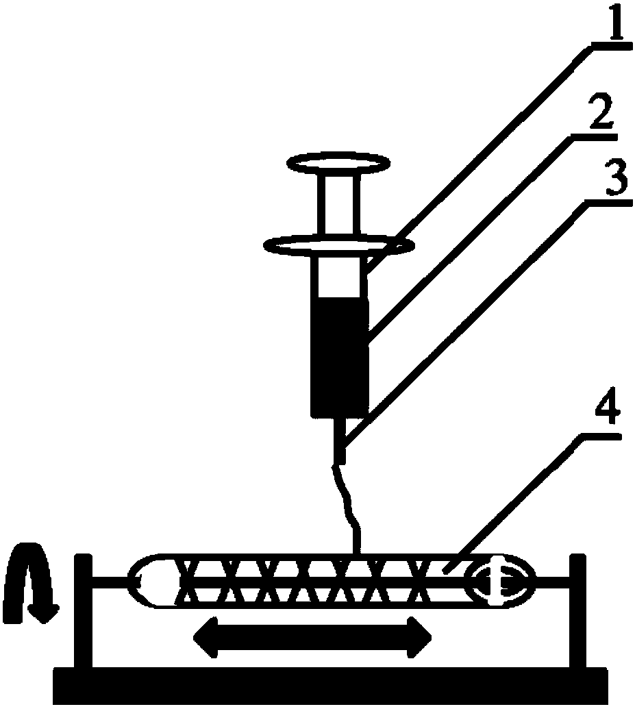
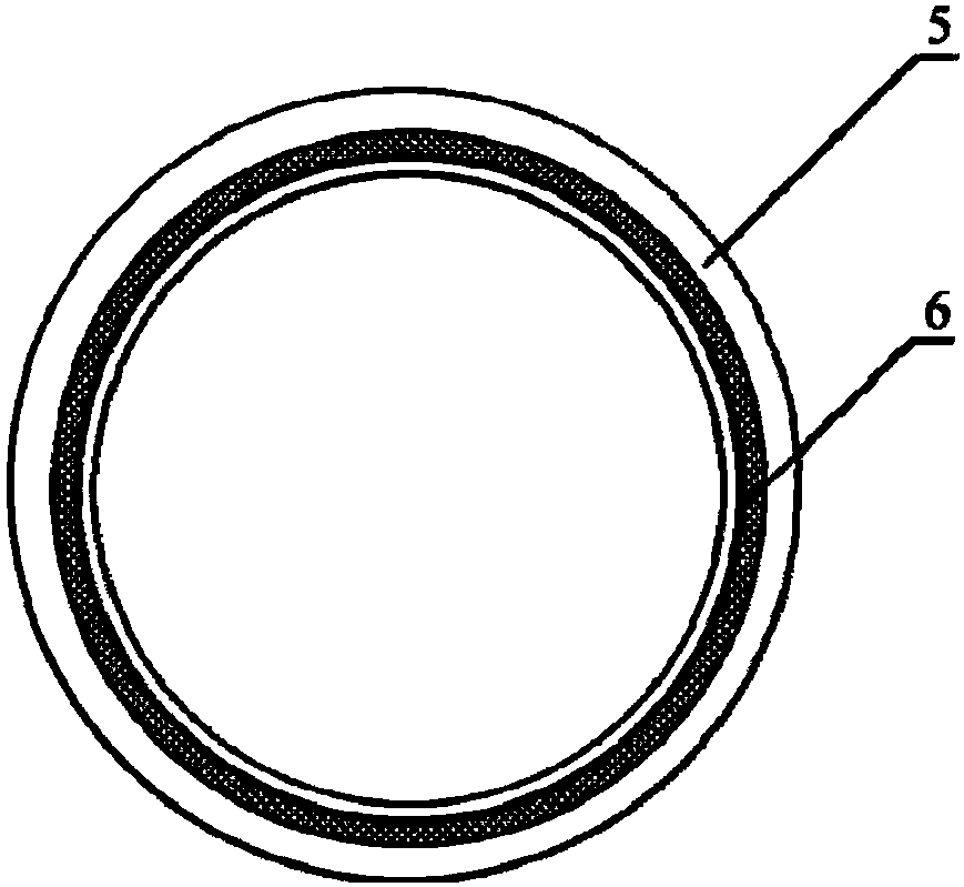
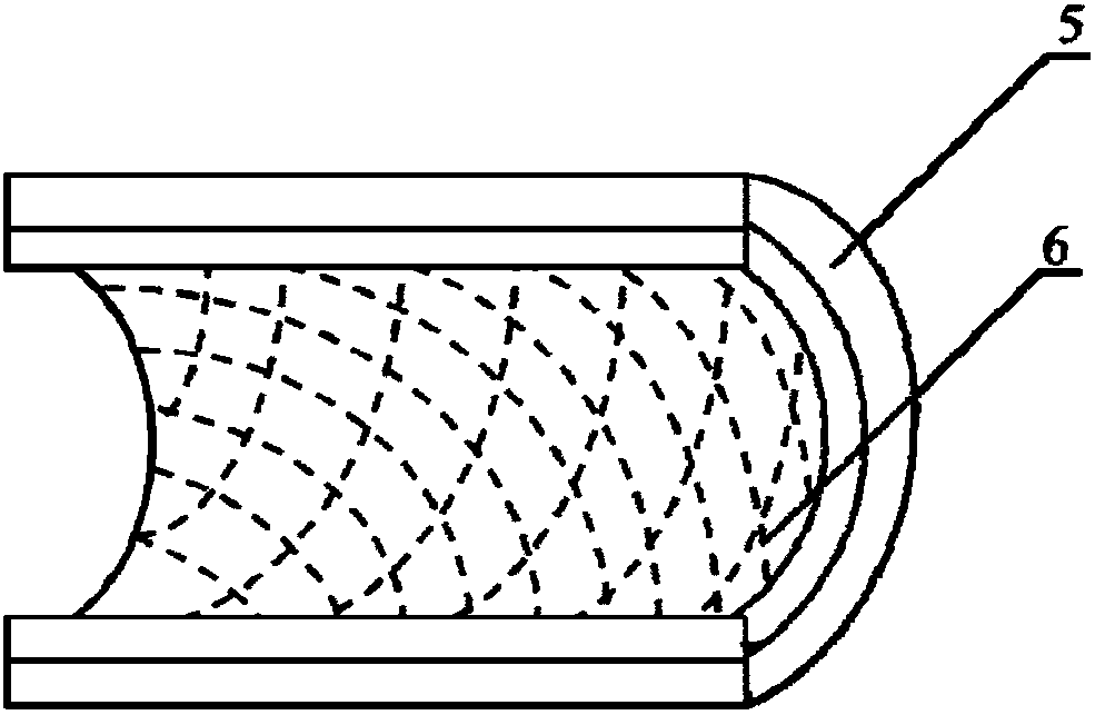
[0033] 1. Prepare the melt spinning fiber skeleton with polycaprolactone (PCL) as spinning fiber material; Take 5.0g by weighing, the number average molecular weight is 80000PCL, place in the closed rust-free steel syringe that thermal melter wraps, in Heat at 200°C for 1h. Put a medical silicone tube with an outer diameter of 2mm and an inner diameter of 1mm on a stainless steel rod with a diameter of 1mm and connect it to the rotating motor. The speed is set to 300r / min, and the translation speed is set to 10mm / s. The receiving output is 45 times, and the composite mandrel of PCL melt-spun fiber and medical silicone tube with a fiber angle of 45° is prepared. The diameter of the PCL melt-spun fiber is 60 μm; the wall of the fiber skeleton tubular structure is 250 μm. In the ice-water mixture, take it out after 5s and let it dry for later use.

[0034] 2. Preparation of engineered arterial vessels in vivo: After anesthetizing the rat, prepare the skin on the back, cut an o...

Embodiment 2

[0039] Embodiment 2. The difference between embodiment 2 and embodiment 1 is that the preparation method of the melt-spun fiber skeleton is different;

[0040] Weigh 5.0 g of PCL with a number average molecular weight of 80,000, place it in a closed stainless steel syringe wrapped in a fuser, and heat it at 200 °C for 1 h. Put a medical silicone tube with an outer diameter of 2mm and an inner diameter of 1mm on a stainless steel rod with a diameter of 1mm and connect it to the rotating motor. The distance between the syringe needle and the receiver is 2mm, the flow rate of the PCL melt is 0.1ml / h, and the rotation of the receiving rod The speed is set to 300r / min, and the translation speed is set to 5mm / s. The receiving output is 200 times, and the composite mandrel of PCL melt-spun fiber and medical silicone tube with a fiber angle of 30° is prepared. The diameter of the spun fiber is 5 μm; the wall thickness of the tubular structure of the prepared PCL fiber skeleton is 250...

Embodiment 3

[0041] Embodiment 3. The difference between embodiment 3 and embodiment 1 is only different in the preparation method of the melt spinning fiber skeleton;

[0042] Weigh 5.0 g of PGA with a number average molecular weight of 100,000, place it in a closed stainless steel syringe wrapped in a fuser, and heat at 300°C for 1 hour. Put a medical silicone tube with an outer diameter of 2mm and an inner diameter of 1mm on a stainless steel rod with a diameter of 1mm and connect it to the rotating motor. The speed is set to 12r / min, and the translation speed is set to 50mm / s. The receiving output is 60 times, and the composite mandrel of PGA melt-spun fiber and medical silicone tube with a fiber angle of 130° is prepared. The diameter of the spun fiber is 50 μm; the wall thickness of the fiber skeleton tubular structure is 300 μm; after the spinning is completed, the silicone tube is withdrawn from the stainless steel rod.


PUM

PropertyMeasurementUnit
diameteraaaaaaaaaa
thicknessaaaaaaaaaa
diameteraaaaaaaaaa

Abstract

The invention belongs to the field of medical equipment and in particular relates to a method for constructing an engineered artery blood vessel in vivo by taking melt-spun fibers as a framework. Theframework comprises a cylindrical receiver and a plurality of spun fibers which are wound on the cylindrical receiver; the diameter of the spun fibers is 5 to 200mu m; the spun fibers comprise warp fibers and weft fibers; a longitudinal included angle between the warp fibers and the weft fibers is alpha and the alpha is greater than or equal to 30 degrees and less than or equal to 130 degrees; thewarp fibers and the weft fibers are staggered for a plurality of layers up and down to form a cylindrical structure. According to the method provided by the invention, the finally-obtained melt-spunfiber framework has good mechanical strength, toughness and compliance through adjusting the diameter of the spun fibers, a winding angle and the winding density, and is suitable for surgical suturingoperation; the framework has good elasticity and can be shrunk or expanded along changes of blood pressure in an application process, the smoothness of the blood vessel is kept, the binary restenosisis reduced and arterial aneurysm is prevented from being formed.

Description

technical field [0001] The invention belongs to the field of medical equipment, and in particular relates to a method for constructing an engineered arterial vessel in vivo using melt-spun fibers as a skeleton. Background technique [0002] Cardiovascular disease and peripheral vascular disease are among the diseases with the highest morbidity and mortality worldwide. It is estimated that by 2030, there will be as many as 23.3 million deaths related to cardiovascular disease. When the blood vessels are less narrow, treatments include angioplasty and stenting. But for highly narrowed or even blocked blood vessels, vessel bypass surgery is necessary, which has reached about 400,000 cases per year in the United States alone. Autologous blood vessels, the gold standard for vascular grafts, such as the saphenous vein and internal mammary artery, are commonly used in arterial bypass surgery. However, the use of autologous blood vessels requires a second operation, which not onl...

Claims


Application Information

Patent Timeline
Patent Type & Authority Applications(China)
IPC IPC(8): A61L27/50A61L27/18D04H1/76D01D5/08A61F2/06
CPCA61L27/507A61F2/06A61L27/18A61L27/50D01D5/08D04H1/76C08L67/04
Inventor 王恺张秋影孔德领朱美峰韩晶黄瑞
Owner NANKAI UNIV
Why Eureka
Try Eureka