
Brand-new nuclear magnetic resonance echo mechanism based magnetic resonance imaging method

A magnetic resonance imaging and nuclear magnetic resonance technology, which is used in magnetic resonance measurement, measurement of magnetic variables, and measurement of magnetic variables. It can solve the problems of gradient echo being unsuitable for diffusion imaging and signal attenuation, and achieve accurate and reliable quantitative measurement methods. , Reduce the signal relaxation attenuation, the effect of broad application prospects

Active Publication Date: 2018-06-22
BEIJING HANSHI MEDICAL TECH CO LTD
View PDF3 Cites 8 Cited by
  • Summary
  • Abstract
  • Description
  • Claims
  • Application Information

AI Technical Summary

Problems solved by technology

normally: Therefore, gradient echo is not suitable for diffusion imaging; spin echo is currently the most used in clinical imaging sequences, but under long diffusion time 2 Relaxation also causes the signal to attenuate a lot
The stimulated echo is due to a period of t m time to T 1 Relaxation can reduce the attenuation of the signal, but it can only gather half of the spin signal, so it is not better than spin echo in any case, but only when the When , the stimulated echo is stronger than the spin echo

Method used


Image

  • Brand-new nuclear magnetic resonance echo mechanism based magnetic resonance imaging method
  • Brand-new nuclear magnetic resonance echo mechanism based magnetic resonance imaging method
  • Brand-new nuclear magnetic resonance echo mechanism based magnetic resonance imaging method

Examples


Embodiment 1

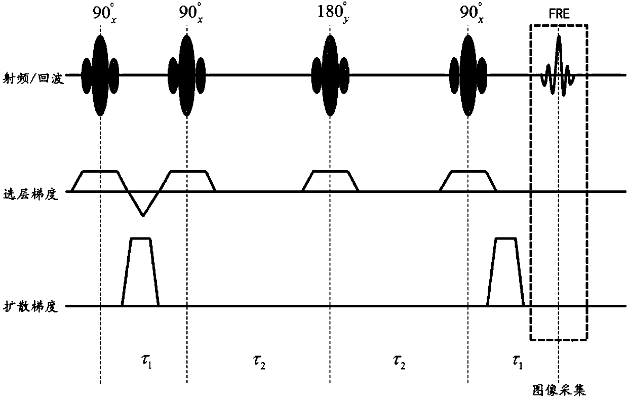
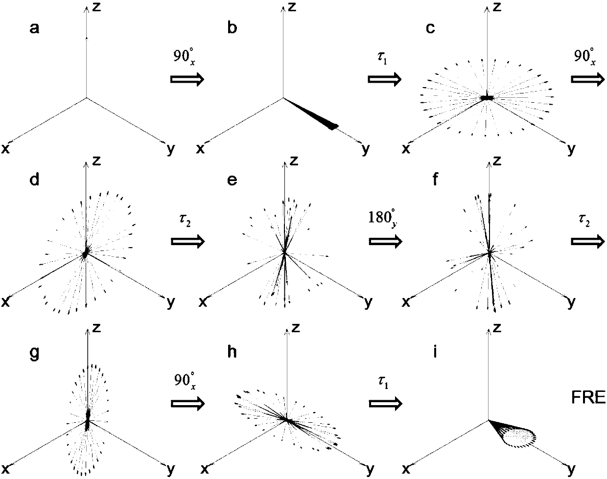
[0045] Such as Figure 1~2 As shown, the present embodiment provides a magnetic resonance imaging method based on a new nuclear magnetic resonance echo mechanism, including the following steps:

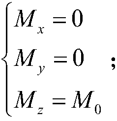
[0046] S1: if figure 2 As shown in a, the initial state spin magnetization vector M 0 All along the +z axis, one of the spins whose off-resonance frequency is ω is selected for tracking observation, assuming that its Larmor precession frequency remains unchanged throughout the whole process, and the relaxation and precession during the RF pulse flipping process are ignored. At this moment, the three components of the spin magnetization vector are:

[0047]

[0048] Excite all the spin magnetization vectors to the +y-axis direction by the first radio frequency pulse with a flip angle of θ=90° along the +x axis, load the layer selection gradient at the same time as the radio frequency excitation to achieve layer selective excitation, and after the excitation is completed Loading ...

experiment example

[0070] A cylindrical test tube with a diameter of 5 cm is filled with an aqueous solution of gadopentetate meglumine with a concentration of 2.5 mM, the transverse relaxation time T2 = 75 ms, and the longitudinal relaxation time T1 = 116 ms; the spin evolution period during detection is set to τ 1 = 12ms, τ 2 = 97ms, using spin echo (SE), stimulated echo (STE) and the echo sequence FRE provided by the present invention to measure and image respectively, measure the signal and noise of the three kinds of echo imaging respectively, and calculate the signal-to-noise Compare. The results are shown in Table 1.

[0071] Table 1 The signal-to-noise ratio of three kinds of echo imaging

[0072]

[0073] It can be seen from Table 1 that the signal-to-noise ratio of FRE imaging is significantly higher than that of SE and STE. Therefore, using the echo sequence provided by the present invention to replace spin echo or stimulated echo in diffusion and flow imaging can greatly improv...


PUM


Abstract

The invention discloses a brand-new nuclear magnetic resonance echo mechanism based magnetic resonance imaging method. The method includes: turning dispersion-phase spins to a longitudinal plane, andsubstituting transverse relaxation with longitudinal relaxation to reduce signal relaxation attenuation; combining with a brand-new echo generating mechanism to obtain completely gathered echo on a transverse plane to keep signals as far as possible, so that a signal-to-noise ratio of an imaging sequence is substantially increased. The method has advantages that the defect of respective signal loss of traditional echo sequences (spin echoes and stimulated echoes) is overcome, optimal echo signals are formed, sequence schemes comprehensively superior to the spin echoes and the stimulated echoesare provided for echo sequence applying diffusion imaging and flow imaging, the signal-to-noise ratio of images is increased to obtain high-quality images, and accordingly an accurate and reliable quantitative measurement method is provided for magnetic resonance in bulk diffusion and flow imaging, and a promising application prospect in biological medical research and clinical diagnosis is achieved.

Description

technical field [0001] The invention belongs to the technical field of magnetic resonance imaging, in particular to a magnetic resonance imaging method based on a new nuclear magnetic resonance echo mechanism. Background technique [0002] Magnetic resonance imaging (MRI) is currently the only imaging modality that can nondestructively observe fluid behavior (flow, diffusion) at any depth inside organisms and the human body. Diagnosis based on the fluid state in the body is a supplement and improvement to the traditional diagnosis based on solid phase information (including pathological slices, X-ray / CT / MRI structural images), and is increasingly accepted in clinical practice because of its specificity and sensitivity. Pay attention to. In particular, diffusion imaging represented by ADC and DTI has irreplaceable value in tumor MRI diagnosis, and also plays an important role in basic research and clinical application of brain science. With the continuous improvement of MRI...

Claims


Application Information

Patent Timeline
IPC IPC(8): G01R33/44G01R33/563
CPCG01R33/448G01R33/56308G01R33/56341
Inventor 王莹
Owner BEIJING HANSHI MEDICAL TECH CO LTD
Learn More