Method for building lipopolysaccharide induced piglet liver cell programmed necrosis model
A technology of programmed necrosis and liver cells, which is applied in the field of establishment of lipopolysaccharide-induced programmed necrosis model of piglet liver cells, can solve the problems of cumbersome steps and achieve good repeatability, reliable measurement results, and simple and easy operation methods
- Summary
- Abstract
- Description
- Claims
- Application Information
AI Technical Summary
Problems solved by technology
Method used
Image


Examples
Embodiment 1
[0052] Embodiment 1 Experimental Design and Tissue Sample Collection
[0053] (1) Select 12 Duroc×Landrace×Large White weaned piglets (8.9±0.2kg, 35±1d), and randomly distribute them to 2 treatment groups (control group and treatment group) according to the principle of similar body weight, each treatment group 6 repetitions. The test piglets were raised in a 1.80×1.10m 2 Stainless steel cages were kept at a temperature of 22-25°C during the feeding period, and food and water were provided ad libitum. After feeding for 10 days, the treatment group was injected with 100 μg of lipopolysaccharide (LPS, Escherichia coli serotype O55: B5, purity >99%, Sigma Company) per kg body weight of piglets, and the control group was injected with an equivalent amount of 0.9% normal saline (injection Stop feeding 12h before).
[0054] (2) 4 hours after the test piglets were injected with LPS (treatment group) or normal saline (control group) peritoneally, 10 mL of blood was collected from t...
Embodiment 2
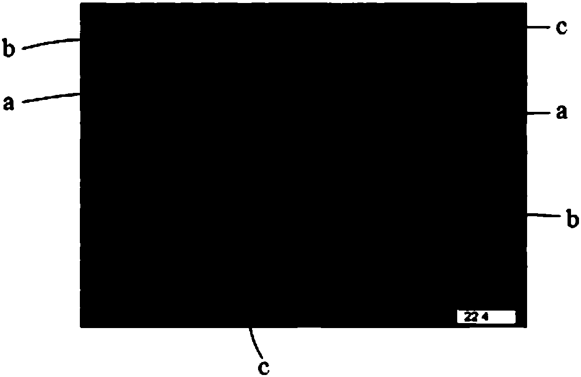
[0056] Example 2 Effect of LPS stimulation on the morphology and structure of piglet liver tissue
[0057] The fresh liver tissue dissected out after slaughter was fixed in 4% paraformaldehyde for more than 24 hours, and the effect of LPS stimulation on the morphology and structure of liver tissue was observed by using a high-definition color pathology graphic report analysis system. The specific method is as follows:
[0058] (1) Take the liver tissue out of the fixative, use a scalpel in the fume hood to flatten the tissue at the target site, put the trimmed tissue and the corresponding label in the dehydration box, and put the dehydration box into the hanging basket Dehydration in the dehydrator with gradient alcohol: 75% alcohol (4h), 85% alcohol (2h), 90% alcohol (2h), 95% alcohol (1h), absolute ethanol I (30min), absolute ethanol II (30min), alcohol benzene (5-10min), xylene I (5-10min), xylene II (5-10min), wax I (1h), wax II (1h), wax III (1h).
[0059] (2) Embed the ...
Embodiment 3
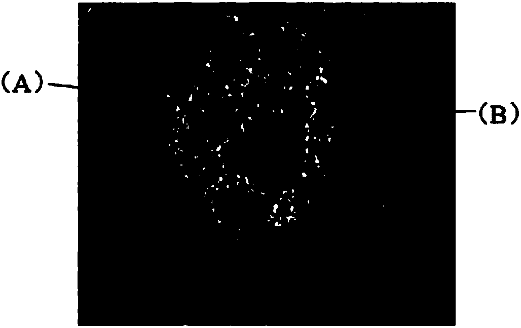
[0064] Example 3 Effect of LPS stimulation on the ultrastructure of piglet liver cells
[0065] The fresh liver tissue dissected after slaughter was fixed in 2.5% glutaraldehyde for more than 4 hours, and Tecnai G was used to 2 20 TWIN transmission electron microscope to observe the effect of LPS stimulation on the ultrastructure of liver cells, the specific method is as follows:
[0066] (1) Cut 2mm 3 The liver samples were added with 2.5% glutaraldehyde, stored and fixed at 4°C for 4 hours, rinsed three times with 0.1M phosphate buffer (pH value 7.4), 15min each time; then pre-cooled at 4°C 1% osmic acid, fixed at 20°C for 2-3 hours; then rinsed with 0.1M phosphate buffer (pH 7.4) for 3 times, 15 minutes each time.
[0067] (2) Dehydrate the liver sample treated in step (1) through graded alcohol (30%, 50%, 70%, 80%, 85%, 90%, 95%, 100% twice), each time for 15-20min . Then the dehydrated liver tissue was infiltrated in a 37°C incubator. The infiltrating agent was aceton...
PUM
| Property | Measurement | Unit |
|---|---|---|
| Weight | aaaaa | aaaaa |
Abstract
Description
Claims
Application Information
 - R&D
- Intellectual Property
- Life Sciences
- Materials
- Tech Scout
- Unparalleled Data Quality
- Higher Quality Content
- 60% Fewer Hallucinations
Browse by: Latest US Patents, China's latest patents, Technical Efficacy Thesaurus, Application Domain, Technology Topic, Popular Technical Reports.
© 2025 PatSnap. All rights reserved.Legal|Privacy policy|Modern Slavery Act Transparency Statement|Sitemap|About US| Contact US: help@patsnap.com