Human pituitary adenoma cell strain and purpose thereof
A technology for pituitary adenomas and cell lines, which is applied in the field of tumor biology and can solve the problems of difficult transfer and non-humanized pituitary tumor cells.
- Summary
- Abstract
- Description
- Claims
- Application Information
AI Technical Summary
Problems solved by technology
Method used
Image
Examples
Embodiment 1
[0041] Embodiment 1: Acquisition and cultivation of human pituitary adenoma cell line
[0042] 1. HPA1446 primary cell culture
[0043] 1. Source of biological material
[0044] The cell line of the invention is obtained by separating and culturing the pituitary tumor tissue of a non-functioning pituitary tumor patient. The tumor tissue was obtained from a surgical specimen of a patient in the Neurosurgery Center of Beijing Tiantan Hospital Affiliated to Capital Medical University. The pathological diagnosis was a non-functional pituitary adenoma. Under the microscope, the tumor cells were densely distributed, with unclear membrane boundaries and inconsistent nuclei in size and shape. , nucleoli and binuclear tumor cells can be seen.
[0045] 2. Isolation and culture method
[0046] Wash the tissue block twice with PBS solution (1:1) containing red solution (purchased from Gibco) in a sterile ultra-clean bench, remove blood vessels and necrotic tissue, and cut the tissue bl...
Embodiment 2
[0065] Embodiment 2: Determination of cell growth curve
[0066] 1. Method:
[0067] The primary pituitary adenoma cells of Example 1 and the immortalized pituitary adenoma cells were adjusted to a cell concentration of 5×10 cells after the cells were overgrown. 4 cells / mL, inoculate primary pituitary tumor cells and immortalized pituitary tumor cells in 96-well culture plate, 100 μl per well, 37°C, 5% CO 2 Cultivate in an incubator; add 10 μl of MTT to each well after culturing for 12h, 24h, 36h, 48h, and 72h respectively, at 37°C, 5% CO 2 Cultivate in the incubator and incubate for 3-4 hours in the dark; absorb the liquid in the well, add 200 μl of DMSO, and shake on the shaker at room temperature for 10 minutes; measure the OD value at the same time point with a wavelength of 492nm on a microplate reader, and use the measured OD value analyze.
[0068] 2. Results:
[0069] Such as figure 1 As shown, the immortalized pituitary tumor cells entered the growth stationary p...
Embodiment 3
[0070] Embodiment 3: cell morphology detection
[0071] 1. Method:
[0072] 1. Observing the cell line HPA1446 (preservation number is CGMCC No. 12672) by phase contrast microscope.
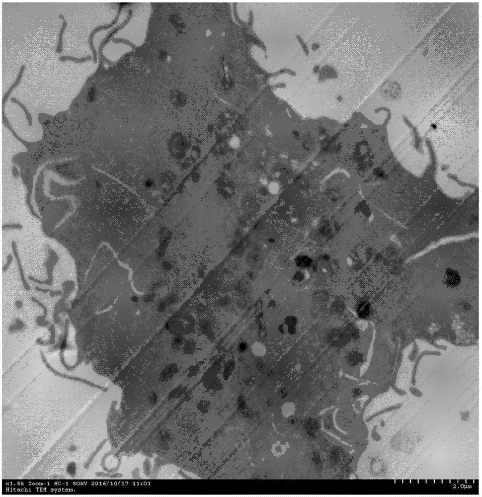
[0073] 2. Observing the ultrastructure of cell line HPA1446 (preservation number is CGMCC No.12672) by transmission electron microscope:
[0074] When the cells grow to 90% confluence, discard the culture medium, add 3ml 0.1MPBS (pH value 7.4), scrape the cells with a rubber cell scraper, collect the cells, centrifuge (1000rpm / min, 10 minutes), discard the supernatant, Aldehyde / osmium tetroxide pre-fixed for 2 hours, washed 3 times with 0.1M PBS (pH 7.4), 10 minutes each time. Then the specimens were post-fixed, dehydrated, soaked, embedded, cut into ultra-thin sections and stained with heavy metals (completed by the electron microscope room of Beijing Institute of Neurosurgery).
[0075] 2. Results:
[0076] 1. Growth characteristics of the cell line HPA1446 (preservation number: CGMCC No.12...
PUM
 Abstract
Description
Claims
Application Information
 - R&D
- Intellectual Property
- Life Sciences
- Materials
- Tech Scout
- Unparalleled Data Quality
- Higher Quality Content
- 60% Fewer Hallucinations
Browse by: Latest US Patents, China's latest patents, Technical Efficacy Thesaurus, Application Domain, Technology Topic, Popular Technical Reports.
© 2025 PatSnap. All rights reserved.Legal|Privacy policy|Modern Slavery Act Transparency Statement|Sitemap|About US| Contact US: help@patsnap.com


