DNA silver nano-clusters and in-situ synthesis method and application of DNA silver nano-clusters in cells
A technology for in situ synthesis of silver nanoclusters, applied in the field of DNA silver nanoclusters, can solve the problems of inability to monitor telomerase activity in real time for a long time and limit the residence time of probes.
- Summary
- Abstract
- Description
- Claims
- Application Information
AI Technical Summary
Problems solved by technology
Method used
Image
Examples
Embodiment 1
[0089] Example 1 In situ synthesis of endogenous DNA silver nanoclusters in cells
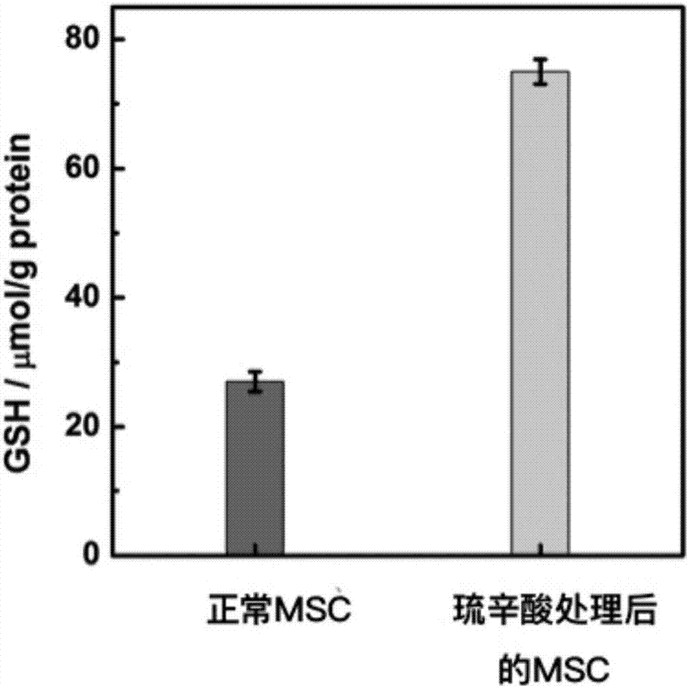
[0090] (1) The embodiment up-regulates the intracellular GSH level:
[0091] Cells were cultured in a 25mL cell culture flask, and when the cell coverage reached about 80%, 10 μL of 5 μM α-lipoic acid (α-lipoic acid) was added and co-incubated for 24 hours at 37 degrees Celsius and 5% CO2, and then slowly poured out the culture solution and replaced with fresh medium.
[0092] The above cells were washed once with PBS, collected by centrifugation, and the supernatant was aspirated. Add a protein removal reagent 3 times the volume of the cell pellet, and then use liquid nitrogen and a 37°C water bath to freeze and thaw the sample twice to break up the cells. After 5 minutes at 4°C or in an ice bath, centrifuge at 10,000 g for 10 minutes at 4°C. The supernatant was taken to determine the content of glutathione with DTNB kit.
[0093] (2) Transfection DNA:
[0094] Take the above-mentioned ce...
Embodiment 2 Embodiment 1
[0098] Example 2 Example 1 The DNA silver nanocluster probe synthesized in situ is used for long-term monitoring of stem cell proliferation
[0099] Changes in telomerase activity during differentiation.
[0100] (1) In situ monitoring of telomerase activity changes in human umbilical cord mesenchymal stem cells during proliferation:
[0101] The first-generation human umbilical cord mesenchymal stem cells treated in Example 1 were cultured in a cell culture box. At each passaging, take part of the plate. The corresponding samples were observed with a fluorescent confocal microscope, excited at a wavelength of 488 nm, and co-fluorescent imaging was observed with an objective lens multiple of 63X. And use the software to calculate the average fluorescence intensity in each cell, and compare the relative activity of telomerase in each generation of mesenchymal stem cells.
[0102] The method for calculating the relative activity of telomerase is to use the average fluorescenc...
Embodiment 3
[0106] Embodiment 3 in vitro verification experiment
[0107] In order to evaluate the accuracy of the DNA silver nanocluster probe synthesized in situ in Example 1 of the present invention for long-term monitoring of changes in telomerase activity in the process of stem cell proliferation and differentiation, each generation of mesenchymal stem cells was lysed and telomeres were extracted. Enzymes The telomerase activity of each generation of cells was detected using an ELISA kit. In addition, neural stem cells differentiated from mesenchymal stem cells of each generation were lysed and telomerase was extracted, and the telomerase activity of cells of each generation was detected using an ELISA kit.
[0108] Figure 9 a is a series of cells used in the verification experiment of Example 3. Figure 9 b is the result of lysing and extracting telomerase from each generation of mesenchymal stem cells, and detecting it with an ELISA kit. It can be seen from the figure that the ...
PUM
| Property | Measurement | Unit |
|---|---|---|
| height | aaaaa | aaaaa |
| particle diameter | aaaaa | aaaaa |
| lattice spacing | aaaaa | aaaaa |
Abstract
Description
Claims
Application Information
 - R&D
- Intellectual Property
- Life Sciences
- Materials
- Tech Scout
- Unparalleled Data Quality
- Higher Quality Content
- 60% Fewer Hallucinations
Browse by: Latest US Patents, China's latest patents, Technical Efficacy Thesaurus, Application Domain, Technology Topic, Popular Technical Reports.
© 2025 PatSnap. All rights reserved.Legal|Privacy policy|Modern Slavery Act Transparency Statement|Sitemap|About US| Contact US: help@patsnap.com


