An aptamer for detecting lactoferrin content and its application
A technology for lactoferrin and protein content, which is used in measurement devices, material excitation analysis, fluorescence/phosphorescence, etc., can solve the problems of complex pretreatment process, low sample recovery rate, poor stability, etc., and achieve high sensitivity and anti-interference. Strong, avoid false positive signal effect
- Summary
- Abstract
- Description
- Claims
- Application Information
AI Technical Summary
Problems solved by technology
Method used
Image
Examples
Embodiment 1
[0023] Example 1: Lactoferrin aptamer screening
[0024] The specific steps of lactoferrin aptamer screening, including chip preparation, positive and negative screening process, PCR amplification and other detailed processes, are as follows, wherein:
[0025] Library: 5′-TAMRA-GACAGGCAGGACACCGTAAC-N40-CTGCTACCTCCCTCCTCTTC-3′
[0026] TARMA modified forward primer: 5′-TARMA-GACAGGCAGGACACCGTAAC-3′
[0027] Biotinylated backward primer: 5′Biotin-GAAGAGGAGGGAGGTAGCAG-3′
[0028] (1) Chip preparation: First, use the combination of photolithography mask and chemical etching to make a microfluidic channel template as a PDMS channel to make a mold; then weigh the PDMS prepolymer with a mass ratio of 10:1 and solidify The reagents were fully mixed, vacuumed, poured onto the microfluidic channel template and cured to obtain a PDMS microfluidic channel; use a spotting instrument to spot a lactoferrin and negative protein microarray with a concentration of 5 mg / mL on the glass substra...
Embodiment 2
[0035]Embodiment 2: Fluorescence polarization method detects lactoferrin standard sample:
[0036] (1) In 10 μL, 250 nM FITC (fluorescein isothiocyanate) labeled aptamer N2 (base sequence: AGGCAGGACACCGTAACCGGTGCATCTATGGCTACTAGCTTTTCCTGCCT) solution, add 100 μL of lactoferrin standard sample with a concentration of 25 μg / mL, fully Mix well.
[0037] (2) The mixture in step (1) was placed in a 96 microwell plate and reacted at 37 °C for 15 min, and scanned directly with a BioTeK microplate reader with an excitation wavelength of 480 nm and an emission wavelength of 528 nm.
Embodiment 3
[0038] Embodiment 3: optimization of reaction time of fluorescence polarization method
[0039] (1) In 10 μL, 250 nM FITC (fluorescein isothiocyanate) labeled aptamer N2 (base sequence: AGGCAGGACACCGTAACCGGTGCATCTATGGCTACTAGCTTTTCCTGCCT) solution, add 100 μL of lactoferrin standard sample with a concentration of 25 μg / mL, fully Mix well.
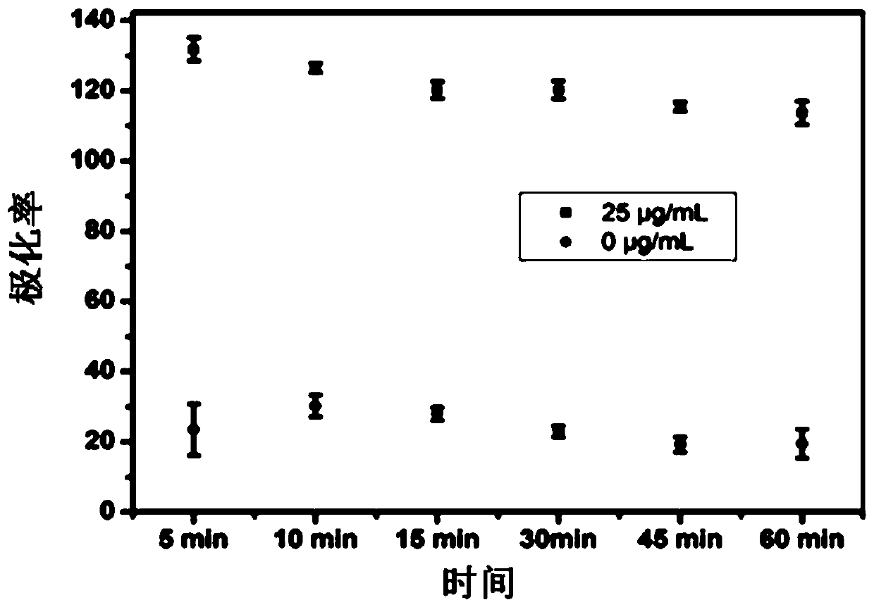
[0040] (2) Put the mixture in step (1) in a 96 microwell plate and react at 37 °C for 5 min, 10 min, 15 min, 20 min, 25 min, 30 min, 35 min, 40 min, 45 min respectively , 50 min, 55 min, 60 min, directly scanned with a BioTeK microplate reader, the excitation wavelength is 480 nm, and the emission wavelength is 528 nm.
[0041] figure 1 It is the graph of the fluorescence polarization signal and the fluorescence polarization signal in the blank solution under different time conditions.
PUM
| Property | Measurement | Unit |
|---|---|---|
| concentration | aaaaa | aaaaa |
Abstract
Description
Claims
Application Information
 - R&D
- Intellectual Property
- Life Sciences
- Materials
- Tech Scout
- Unparalleled Data Quality
- Higher Quality Content
- 60% Fewer Hallucinations
Browse by: Latest US Patents, China's latest patents, Technical Efficacy Thesaurus, Application Domain, Technology Topic, Popular Technical Reports.
© 2025 PatSnap. All rights reserved.Legal|Privacy policy|Modern Slavery Act Transparency Statement|Sitemap|About US| Contact US: help@patsnap.com


