Probe for clearly distinguishing cell membrane-lipid raft microdomain from non-lipid-raft microdomain by using two fluorescence colors and simultaneously imaging microdomains and application of probe

A technology of fluorescent probes and cell membranes, applied in the field of fluorescent probes, can solve the problems of limited application and difficulty in distinguishing two types of micro-regions, and achieve the effects of convenient operation, broad commercialization prospects, and good biocompatibility

Active Publication Date: 2017-05-10
SHANDONG UNIV
View PDF6 Cites 15 Cited by
  • Summary
  • Abstract
  • Description
  • Claims
  • Application Information

AI Technical Summary

Problems solved by technology

However, laurdan has obvious disadvantages: firstly, the difference between the two fluorescent wavelengths is small, only 50nm, which makes it difficult to clearly distinguish the two microdomains on the cell membrane; secondly, it will quickly internalize into the cell, which is also serious Limits its use in imaging research

Method used


Image

  • Probe for clearly distinguishing cell membrane-lipid raft microdomain from non-lipid-raft microdomain by using two fluorescence colors and simultaneously imaging microdomains and application of probe
  • Probe for clearly distinguishing cell membrane-lipid raft microdomain from non-lipid-raft microdomain by using two fluorescence colors and simultaneously imaging microdomains and application of probe
  • Probe for clearly distinguishing cell membrane-lipid raft microdomain from non-lipid-raft microdomain by using two fluorescence colors and simultaneously imaging microdomains and application of probe

Examples


Embodiment 1

[0025] Synthesis of 4,4-dibromo-2-nitrobiphenyl (1)

[0026] 10 g of p-dibromobiphenyl was dissolved in 120 mL of glacial acetic acid, then the solution was stirred and heated to 100 °C. Add 40mL of fuming nitric acid, and react the system for 30 minutes. After the reaction was completed, the solution was cooled to room temperature, and the crude product was obtained by filtration. The pure product can be obtained by recrystallization from ethanol with a yield of 91%.

[0027] 1 H NMR (300MHz, CDCl 3 ), δ (ppm): 8.03 (d, J = 1.8Hz, 1H), 7.76 (dd, J 1 =8.1Hz,J 2 =1.8Hz, 1H), 7.54–7.59(m, 2H), 7.31(s, 1H), 7.14–7.18(m, 2H).

[0028] Synthesis of 2,7-Dibromocarbazole (2)

[0029] 7.8 g of compound 1 was dissolved in 30 mL of triethyl phosphite and stirred evenly. Under the protection of nitrogen, the above system was heated to 150° C. and reacted for 24 hours. After the excess solvent was distilled off under reduced pressure, the residue was separated and purified by col...

Embodiment 2

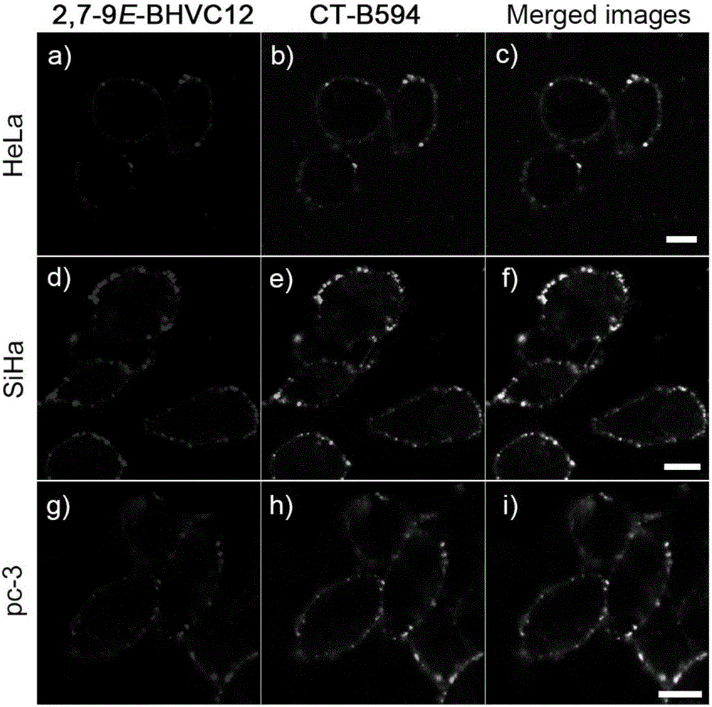
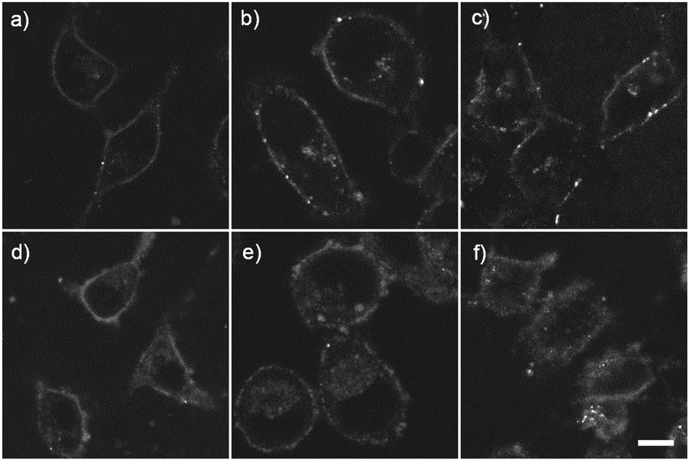
[0041] Culture of SiHa, HeLa and pc-3 cells

[0042] All cell lines were stored at 37°C, 5% CO 2 cultured in a saturated humidity incubator. SiHa and HeLa cell lines were cultured adherently in H-DMEM medium containing 10% fetal bovine serum (containing 1% double antibody). The pc-3 cell line was adhered and cultured in 1640 medium containing 10% fetal bovine serum. After the cells grow to the logarithmic phase, culture the slices: ① Soak the coverslips in absolute ethanol for 30 minutes, dry them with an alcohol lamp, and place them in a disposable 35mm culture dish; ② Wash the cells in the 100mL cell bottle with PBS. Three times, digest with 1mL 0.25% trypsin for 3-5min, pour out the medium carefully, add a small amount of fresh medium and pipette evenly, after counting the cells, leave cells with a suitable density, add the medium to the required volume ( The final concentration of control cells was 1×10 4 ), inoculated into a petri dish containing a coverslip, and plac...

Embodiment 3

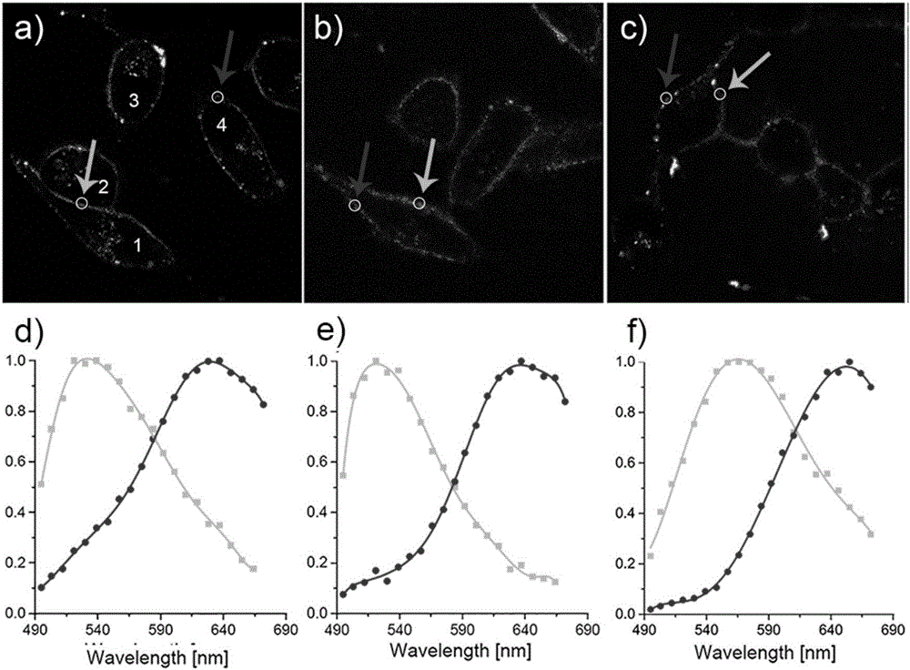
[0044] 2,7-9E-BHVC12 staining of SiHa, HeLa and pc-3 cells and real color imaging

[0045] Firstly, the probe 2,7-9E-BHVC12 was prepared into a DMSO mother solution with a concentration of 5 mM. During the staining experiment, 4 μL of the above mother solution was taken out and added to 1 mL of PBS buffer solution, and the staining solution was made by shaking evenly. The inoculated cell slides were washed three times with PBS, then placed in staining solution for staining, and placed in CO 2 Stain in the incubator for 20min. The staining solution was sucked out, and the stained cell slides were washed with PBS three times, and the cell growth side was covered on the glass slide, and imaged and observed under a laser scanning confocal fluorescence microscope. The full-color fluorescence images were acquired with the Lambda spectral imaging module of a Zeiss confocal microscope LSM780. The module uses a 488nm laser as a light source to excite, and collects fluorescence in the...


PUM


Abstract

The invention discloses a single fluorescent probe for clearly distinguishing a cell membrane-lipid raft microdomain from a non-lipid-raft microdomain by using two fluorescence colors in a real-time in-situ manner and simultaneously imaging the microdomains. The fluorescent probe is a compound with a structure shown as a formula (I), wherein R1 expresses 2-ethoxyethyl, aminoalkyl, hydroxyalkyl or alkyl; R2 and R3 express alkyl. The invention also discloses application of the fluorescent probe in marking the lipid raft microdomain and the non-lipid-raft microdomain on a cell membrane of a living cell by using red and green fluorescence colors in the real-time in-situ manner. The probe can mark the lipid raft microdomain by using red fluorescence, and marks the non-lipid-raft microdomain by using green fluorescence at the same time, thereby realizing simultaneous imaging of the two microdomains. Compared with a current commonly-used similar probe laurdan, the probe disclosed by the invention has the characteristics that spectrum difference of two microdomains is greater, so that clear distinguishing and simultaneous imaging of the two microdomains are realized; besides, the internal problems is improved, so the probe is simpler and more convenient to operate during application and is broad in commercial application prospect.

Description

technical field [0001] The present invention relates to a fluorescent probe, in particular to a single fluorescent probe capable of clearly distinguishing and simultaneously imaging cell membrane lipid rafts and non-lipid raft microregions with two fluorescent colors and its ability to label lipid rafts and non-lipid rafts on the cell membrane of living cells Applications in non-lipid raft microdomains. Background technique [0002] The submicrostructure of cell membranes has attracted extensive attention in recent years. Phospholipids containing saturated fatty acid side chains such as sphingomyelin and cholesterol can form densely arranged lipid raft microdomains, while phospholipids containing unsaturated side chains can form loosely arranged non-lipid raft microdomains. Studies have shown that lipid raft microdomains are closely related to protein cluster formation, signal transduction, cell apoptosis and virus invasion. On the other hand, non-lipid raft domains, due t...

Claims


Application Information

Patent Timeline
IPC IPC(8): C07D401/14C09K11/06G01N21/64
CPCC07D401/14C09K11/06C09K2211/1029G01N21/6486
Inventor 于晓强田明刚何秀全张若瑶
Owner SHANDONG UNIV
Why Eureka
Try Eureka